

Cytotoxic (salen)ruthenium(iii) anticancer complexes exhibit different modes of cell death directed by axial ligands
- PMID: 29147511
- PMCID: PMC5632802
- DOI: 10.1039/c7sc02205k
Cytotoxic (salen)ruthenium(iii) anticancer complexes exhibit different modes of cell death directed by axial ligands
Abstract
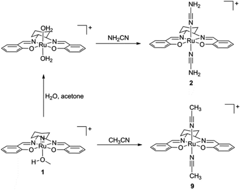
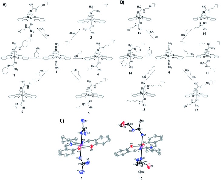
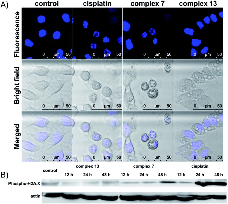
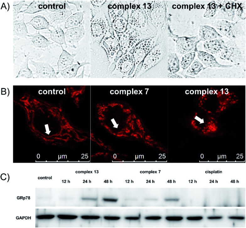
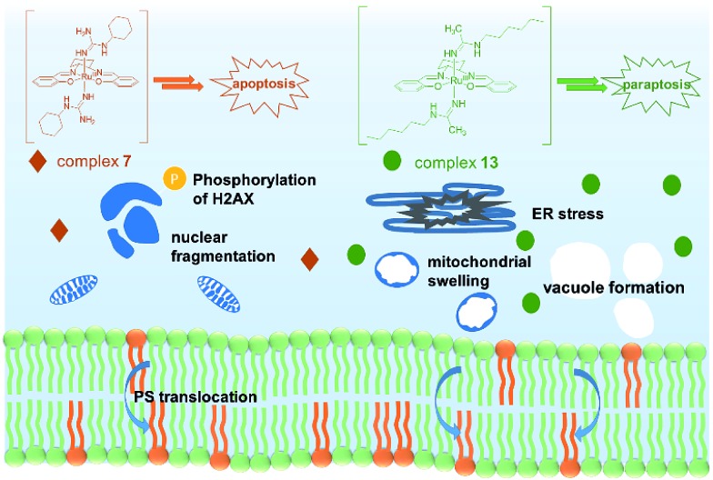
Two novel series of (salen)ruthenium(iii) complexes bearing guanidine and amidine axial ligands were synthesized, characterized, and evaluated for anticancer activity. In vitro cytotoxicity tests demonstrate that these complexes are cytotoxic against various cancer cell lines and the leading complexes have remarkable cancer-cell selectivity. A detailed study of the guanidine complex 7 and the amidine complex 13 reveals two distinguished modes of action. Complex 7 weakly binds to DNA and induces DNA damage, cell cycle arrest, and typical apoptosis pathways in MCF-7 cells. In contrast, complex 13 induces paraptosis-like cell death hallmarked by massive vacuole formation, mitochondrial swelling, and ER stress, resulting in significant cytotoxicity against human breast cancer cells. Our results provide an extraordinary example of tuning the mechanism of action of (salen)ruthenium(iii) anticancer complexes by modifying the structure of the axial ligands.
Figures
References
-
- Brabec V., Kasparkova J. Drug Resist. Updates. 2005;8:131–146. - PubMed
-
- Kelland L. Nat. Rev. Cancer. 2007;7:573–584. - PubMed
-
- Yan Y. K., Melchart M., Habtemariam A., Sadler P. J. Chem. Commun. 2005:4764–4776. - PubMed
-
- Bergamo A., Sava G. Dalton Trans. 2007:1267–1272. - PubMed
-
- Hartinger C. G., Jakupec M. A., Zorbas-Seifried S., Groessl M., Egger A., Berger W., Zorbas H., Dyson P. J., Keppler B. K. Chem. Biodiversity. 2008;5:2140–2155. - PubMed
LinkOut - more resources
Full Text Sources
Other Literature Sources
Molecular Biology Databases