. 2017 Dec 6;7(6):20160127.
doi: 10.1098/rsfs.2016.0127. Epub 2017 Oct 20.

Development of proteolytically stable N-methylated peptide inhibitors of aggregation of the amylin peptide implicated in type 2 diabetes

Affiliations

Development of proteolytically stable N-methylated peptide inhibitors of aggregation of the amylin peptide implicated in type 2 diabetes

Idira Obasse et al. Interface Focus. .

Abstract

Islet amyloid polypeptide, also known as amylin, is the main component of the amyloid deposits present in approximately 90% of people with type 2 diabetes mellitus (T2DM). In this disease, amylin aggregates into multimeric β-pleated sheet structures which cause damage to pancreatic islet β-cells. Inhibitors of early-stage amylin aggregation could therefore provide a disease-modifying treatment for T2DM. In this study, overlapping peptides were designed to target the 'binding' region (RLANFLVHSS, residues 11-20) of human amylin, and their effects on amyloid fibril formation were determined by thioflavin-T assay. The first generation peptides showed less than 50% inhibition of aggregation, but a second generation peptide (H2N-RGANFLVHGR-CONH2) showed strong inhibitory effects on amylin aggregation, and this was confirmed by negative stain electron microscopy. Cytotoxicity studies revealed that this peptide protected human pancreatic 1.4E7 (ECACC 10070102) insulin-secreting cells from the toxic effects of human amylin. Unlike the retro-inverso version of this peptide, which stimulated aggregation, two N-methylated peptides (H2N-RGAmNFmLVmHGR-CONH2 and H2N-RGANmFLmVHmR-CONH2) gave very clear dose-dependent inhibition of fibril formation. These two peptides were also stable against a range of different proteolytic enzymes, and in human plasma. These N-methylated peptides could provide a novel treatment for slowing progression of T2DM.

Keywords: IAPP; N-methylated peptide; amylin; amyloid; diabetes; islet amyloid polypeptide.


Conflict of interest statement

We declare we have no competing interests.

Figures

Figure 1.
Figure 1.
Example of ThT fluorescence curves for human amylin in the presence of different concentrations of an inhibitor (IO4). Data are means for a single experiment carried out in triplicate, with readings taken every 10 min. Amylin alone (at 25 µM) displays a characteristic increase in fluorescence corresponding to the ‘sigmoidal’ and ‘plateau’ phases of amyloid fibril formation, while the addition of the inhibitor, at varying concentrations, has a dose-dependent effect on fibril formation. The buffer control (‘control’) contained neither amylin nor inhibitor.
Figure 2.
Figure 2.
ThT data showing effects of IO1, IO2, IO3, IO4, IO5, IO6 and IO7 peptides on amylin aggregation, after 48 h incubation. All peptides were assayed at 0.6, 2.5, 5, 12.5, 25, 50 and 100 µM concentrations in the presence of 25 µM amylin. Results are means ± s.e.m., n = 3, for a single experiment. Student's t-test was used to establish significance at *p < 0.05, **p < 0.01, or ***p < 0.001, compared to 100% control (amylin alone). (Online version in colour.)
Figure 3.
Figure 3.
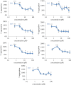
ThT data showing the effects of IO8 and related peptides, as well NFGAILS and NMeG24 NMeI26, on amylin aggregation, after 48 h incubation. (a) IO8 and RI-IO8. (b) N1-IO8 and N2-IO8. (c) NFGAILS (H2N-RGNFGAILSGR-CONH2). (d) NMeG24 NMeI26. All peptides were assayed at 0.6, 2.5, 5, 12.5, 25, 50 and 100 µM in the presence of 25 µM amylin. Results show means ± s.e.m., n = 3, for a single experiment. See electronic supplementary material, figure S2 for the data from three independent experiments. Note the clear dose-dependent effects of the two N-methylated peptides (N1-IO8 and N2-IO8). (Online version in colour.)
Figure 4.
Figure 4.
ThT fluorescence curves for the first 10 h of incubation of amylin in the presence of different concentrations of (a) N1-IO8 and (b) N2-IO8. Data are means for a single experiment carried out in triplicate, with readings taken every 10 min. Amylin alone (at 25 µM) displays a characteristic increase in fluorescence corresponding to the ‘sigmoidal’ and ‘plateau’ phases of amyloid fibril formation (top curve in both cases). In both (a) and (b), the stepwise decrease in the final level of ThT fluorescence in the 10 curves underneath is due to addition of the inhibitors at concentrations of 0.1, 0.3, 0.6, 1, 2.5, 5, 12.5, 25, 50 and 100 µM.
Figure 5.
Figure 5.
Negative stain EM images of amylin incubated in the presence and absence of inhibitors. (a) Sample of amylin (25 µM) incubated for 48 h in PBS at room temperature and stained with phosphotungstic acid (2% w/v). (b) Amylin (25 µM) + IO8 (25 µM); (c) amylin (25 µM) + RI-IO8 (25 µM); (d) amylin (25 µM) + N1-IO8 (25 µM); (e) amylin (25 µM) + N2-IO8 (25 µM); (f) IO8 alone (100 µM); (g) RI-IO8 alone (100 µM); (h) N1-IO8 alone (100 µM); and (i) N1-IO8 alone (100 µM). Scale bar, 100 nm.
Figure 6.
Figure 6.
Reverse-phase HPLC traces showing stability of peptide inhibitors in human plasma. Column (A) IO8; column (B) N1-IO8; column (C) N2-IO8. For each column, the top trace shows elution of the peptide standard without plasma; the middle trace is for 0 h incubation in plasma; and the lower trace is after 48 h incubation in plasma. Note that IO8 is degraded, whereas N1-IO8 and N2-IO8 are more stable.
Figure 7.
Figure 7.
Cytotoxic effect of amylin on human pancreatic 1.4E7 insulin-secreting cells in the presence or absence of inhibitor peptides. (a) Cells were incubated for 24 h with 20 µM or 10 µM human amylin in RPMI-1640 medium, with or without IO8 inhibitor, and viability was measured using the MTS assay. (b) Results for N1-IO8. (c) Results for N2-IO8. In all cases, results show mean ± s.e.m., n = 6. ANOVA followed by Student's t-test established significance at *p < 0.05, **p < 0.01, ***p < 0.001. (Online version in colour.)

References

    1. Dobson CM. 1999. Protein misfolding, evolution and disease. Trends Biochem. Sci. 24, 329–332. (10.1016/S0968-0004(99)01445-0) - DOI - PubMed
    1. Rochet JC, Lansbury PT. 2000. Amyloid fibrillogenesis: themes and variations. Curr. Opin. Struct. Biol. 10, 60–68. (10.1016/S0959-440X(99)00049-4) - DOI - PubMed
    1. Westermark P. et al 2007. A primer of amyloid nomenclature. Amyloid 14, 179–183. (10.1080/13506120701460923) - DOI - PubMed
    1. Westermark P. 2005. Aspects on human amyloid forms and their fibril polypeptides. FEBS Lett. 272, 5942–5949. (10.1111/j.1742-4658.2005.05024.x) - DOI - PubMed
    1. Pepys MB. 2006. Amyloidosis. Annu. Rev. Med. 57, 223–241. (10.1146/annurev.med.57.121304.131243) - DOI - PubMed