. 2017 Jun;2(3):229-240.
doi: 10.1016/j.jacbts.2017.02.004. Epub 2017 Jun 26.

Lipoprotein(a) Associated Molecules are Prominent Components in Plasma and Valve Leaflets in Calcific Aortic Valve Stenosis

Affiliations

Lipoprotein(a) Associated Molecules are Prominent Components in Plasma and Valve Leaflets in Calcific Aortic Valve Stenosis

Michael Torzewski et al. JACC Basic Transl Sci. 2017 Jun.

Abstract

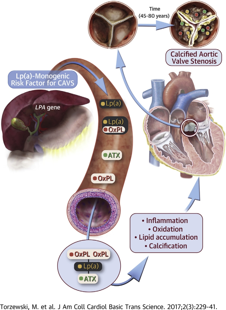
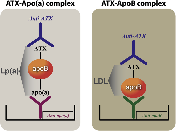
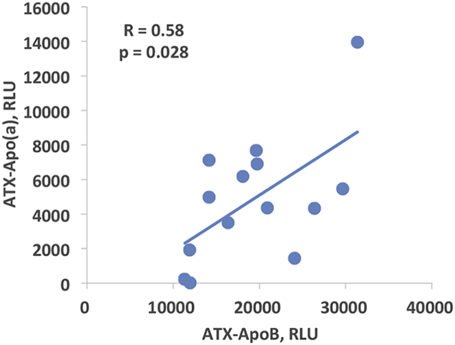
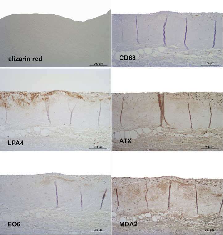
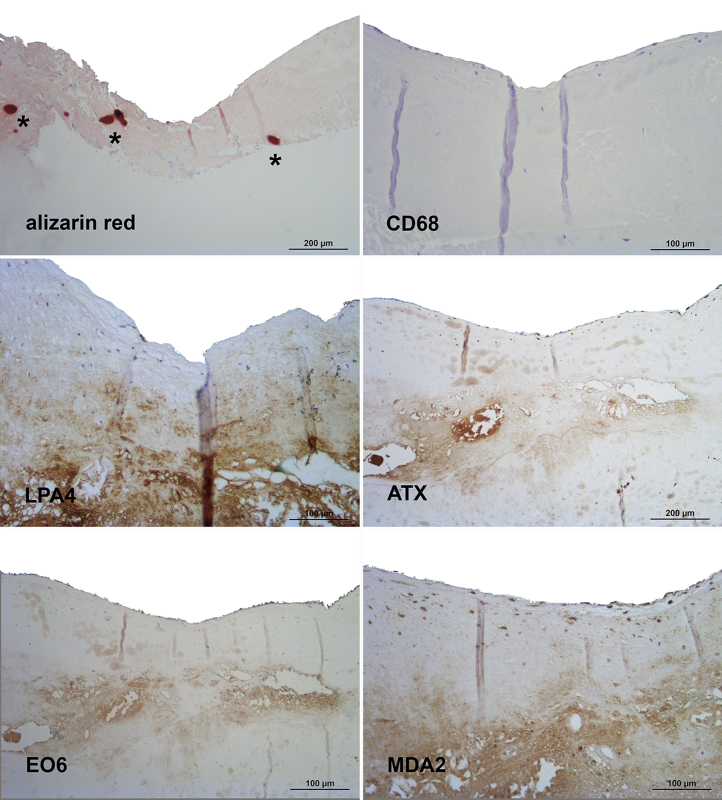
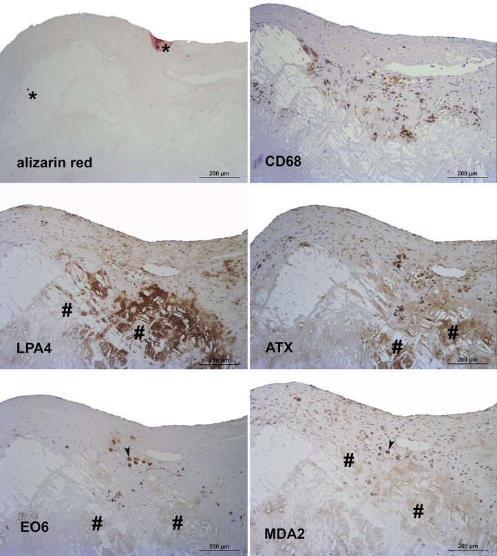
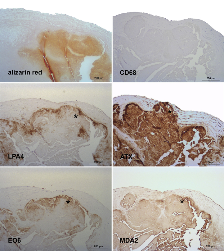
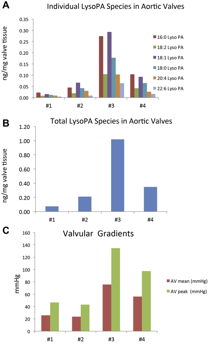
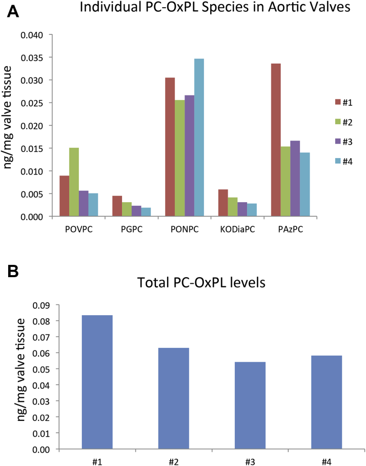
The LPA gene is the only monogenetic risk factor for calcific aortic valve stenosis (CAVS). Oxidized phospholipids (OxPL) and lysophosphatidic acid generated by autotaxin (ATX) from OxPL are pro-inflammatory. Aortic valve leaflets were categorized pathologically from Both ATX-apoB and ATX-apo(a) were measureable in plasma. Lp(a), autotaxin, OxPL and MDA epitopes progressively increased in immunostaining (p<0.001 for all). Six species of OxPL and LysoPA were identified following extraction from valve leaflets. The presence of a constellation of pathologically-linked, Lp(a)-associated molecules in plasma and in aortic valve leaflets of patients with CAVS suggest that Lp(a) is a key etiological factor in CAVS.

Keywords: Lp(a); aortic valve stenosis; autotaxin; inflammation; oxidation-specific epitopes.


Figures

None
Graphical abstract
Figure 1
Figure 1
ELISAs to Measure ATX-Lp(a) and ATX-apoB Methodology of novel chemiluminescent enzyme-linked immunoadsorbent assay (ELISA) to measure autotaxin (ATX)–apolipoprotein(a) (apo[a]) and ATX–apolipoprotein B (apoB) complexes. ApoB-100 and lipoprotein(a) (Lp[a]) lipoproteins were captured from plasma with specific antibodies bound to microtiter well plates. Conditions were established so that the added plasma contains saturating amounts of lipoproteins to be captured. The content of ATX mass on each lipoprotein was then detected with a goat anti-human ATX antibody.
Figure 2
Figure 2
Correlation Between ATX-Apo(a) and ATX-apoB Spearman correlation between plasma levels of ATX-apoB and ATX-apo(a) complexes in patients with mild to moderate calcific aortic valve stenosis. RLU = relative light units; other abbreviations as in Figure 1.
Figure 3
Figure 3
Presence of Apolipoprotein(a), Oxidation-Specific Epitopes, and Autotaxin in Grade 1 of Aortic Valve Disease Sequential sections stained for calcification with alizarin red S and macrophages (CD68) (upper panel), apolipoprotein(a) (LPA4) and autotaxin (ATX) (middle panel) as well as oxidized phospholipids (EO6) and malondialdehyde epitopes (MDA2) (lower panel). Note the lack of calcified areas as well as the predominantly extracellular co-localization of the different antigens. In all panels, the aortic side of the valve is at the top.
Figure 4
Figure 4
Presence of Apolipoprotein(a), Oxidation-Specific Epitopes, and Autotaxin in Grade 2 of Aortic Valve Disease Sequential sections stained for alizarin red S and macrophages (CD68) (upper panel), apolipoprotein (a) (LPA4), and autotaxin (ATX) (middle panel), as well as oxidized phospholipids (EO6) and malondialdehyde epitopes (MDA2) (lower panel). Note the small calcified areas (asterisks), the lack of CD68 staining in this individual lesion (high variation of macrophages staining in aortic valve sclerosis), as well as the predominantly extracellular co-localization of the different antigens. In all panels, the aortic side of the valve is at the top.
Figure 5
Figure 5
Presence of Apolipoprotein(a), Oxidation-Specific Epitopes, and Autotaxin in Grade 3 of Aortic Valve Disease Sequential sections stained for alizarin red S and macrophages (CD68) (upper panel), apolipoprotein (a) (LPA4) and autotaxin (ATX) (middle panel), as well as oxidized phospholipids (EO6) and malondialdehyde epitopes (MDA2) (lower panel). Note the predominance of cholesterol crystals compared to calcified areas (∗) in this individual lesion and co-localization of the different antigens around cholesterol crystal deposits (#). Sometimes additional intracellular oxidized phospholipid staining is also evident (arrowheads). In all panels, the aortic side of the valve is at the top.
Figure 6
Figure 6
Presence of Apolipoprotein(a), Oxidation-Specific Epitopes, and Autotaxin in Grade 4 of Aortic Valve Disease Sequential sections stained for alizarin red S and macrophages (CD68) (upper panel), apolipoprotein (a) (LPA4), and autotaxin (ATX) (middle panel), as well as oxidized phospholipids (EO6) and malondialdehyde epitopes (MDA2) (lower panel). Note the co-localization of the different antigens around and within heavily calcified areas (∗) as well as the lack of CD68 staining in this individual lesion (high variation of macrophages staining in aortic valve sclerosis). In all panels, the aortic side of the valve is at the top.
Figure 7
Figure 7
Presence of LysoPA Species in Valve Leaflets and Relationship to Valve Gradients Prevalence of (A) individual lysophosphatidic acid (LysoPA) species and (B) total LysoPA mass and (C) mean and peak valvular gradients in 4 different stenotic aortic valves. Reverse-phase separation followed by tandem mass spectrometry detection of (A) LysoPA species 16:0, 18:2, 18:1, 18:0, 20:4 and 22:6 in 4 aortic valves and (B) the total LysoPA amounts per milligram of valve tissue extracted. (C) Represents the mean and peak valvular gradients for each of the study valves.
Figure 8
Figure 8
Presence of Individual PC-OxPL Species in Valve Leaflets Liquid chromatography-tandem mass spectrometry analysis of the most abundant fragmented phosphocholine-containing oxidized phospholipid (PC-OxPLs) compounds extracted from human stenotic aortic valves and represented as (A) individual PC-OxPL compounds or (B) as total PC-OxPL levels within each of the valves. Reverse phase separation coupled with tandem mass spectrometry detection was used to detect POVPC (1-palmitoyl-2-[5'-oxo-valeroyl]-sn-glycero-3-phosphocholine), PGPC (1-palmitoyl-2-glutaryl-sn-glycero-3-phosphocholine), PONPC (1-palmitoyl-2-[9'-oxononanoyl]-sn-glycero-3-phosphocholine), KODiA-PC (1-[palmitoyl]-2-[5-keto-6-octene-dioyl]-sn-glycero-3-phosphocholine), and PAzPC (1-palmitoyl-2-azelaoyl-sn-glycero-3-phosphocholine).

References

    1. Rajamannan N.M., Evans F.J., Aikawa E. Calcific aortic valve disease: not simply a degenerative process: a review and agenda for research from the National Heart and Lung and Blood Institute Aortic Stenosis Working Group. Executive summary: calcific aortic valve disease-2011 update. Circulation. 2011;124:1783–1791. - PMC - PubMed
    1. Yutzey K.E., Demer L.L., Body S.C. Calcific aortic valve disease: a consensus summary from the Alliance of Investigators on Calcific Aortic Valve Disease. Arterioscler Thromb Vasc Biol. 2014;34:2387–2393. - PMC - PubMed
    1. Deeb G.M., Reardon M.J., Chetcuti S. 3-Year outcomes in high-risk patients who underwent surgical or transcatheter aortic valve replacement. J Am Coll Cardiol. 2016;67:2565–2574. - PubMed
    1. Capoulade R., Chan K.L., Yeang C. Oxidized phospholipids, lipoprotein(a), and progression of calcific aortic valve stenosis. J Am Coll Cardiol. 2015;66:1236–1246. - PubMed
    1. Kamstrup P.R., Tybjaerg-Hansen A., Nordestgaard B.G. Elevated lipoprotein(a) and risk of aortic valve stenosis in the general population. J Am Coll Cardiol. 2014;63:470–477. - PubMed