Review
. 2018 Apr;10(2):641-658.
doi: 10.1007/s12551-017-0336-9. Epub 2017 Nov 16.

Artificial bio-nanomachines based on protein needles derived from bacteriophage T4

Affiliations
Review

Artificial bio-nanomachines based on protein needles derived from bacteriophage T4

Hiroshi Inaba et al. Biophys Rev. 2018 Apr.

Abstract

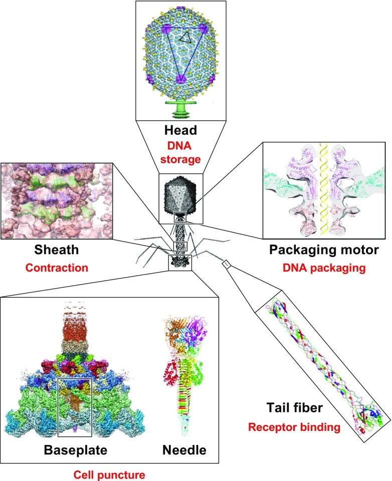
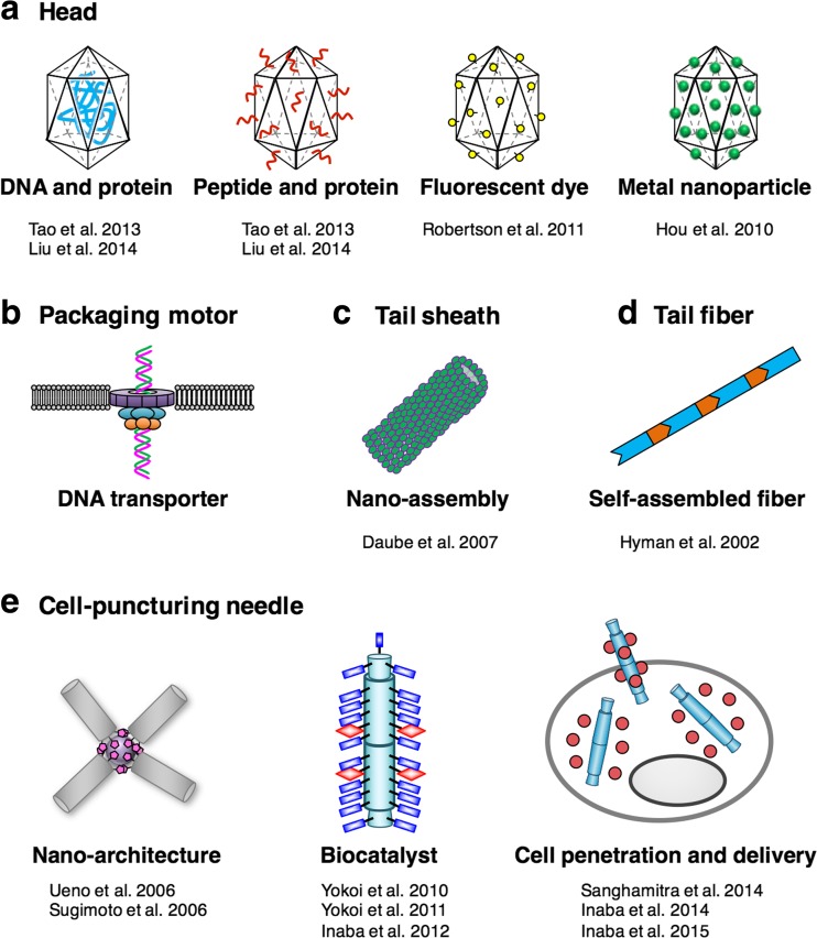
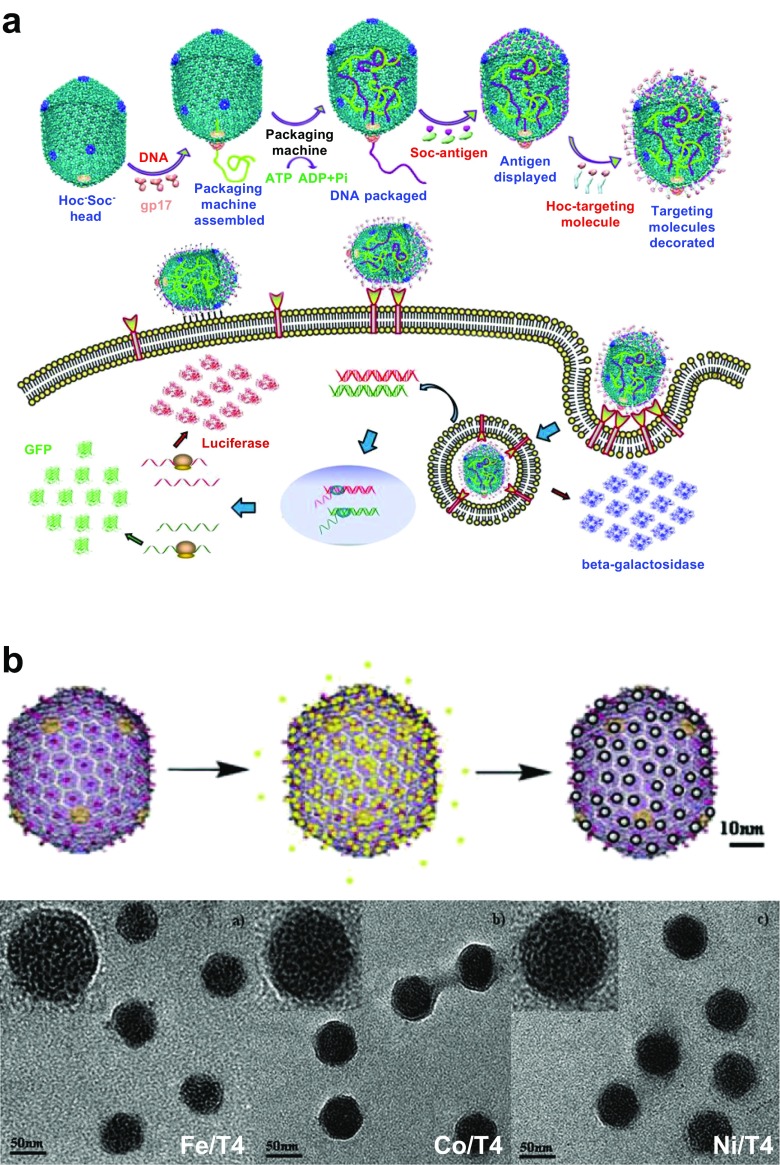
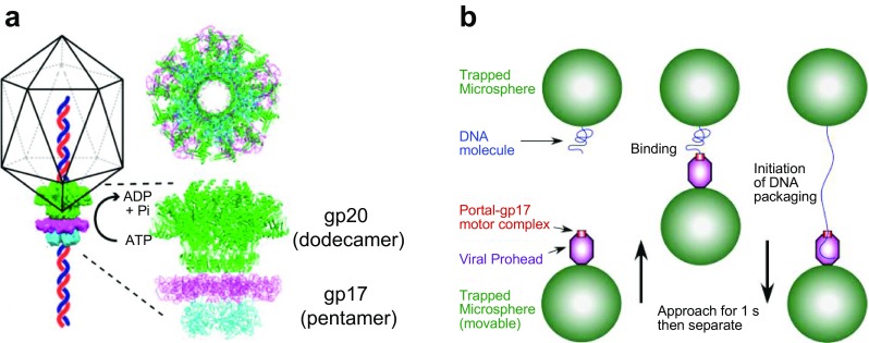
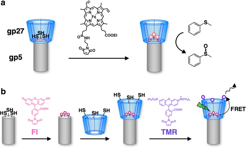
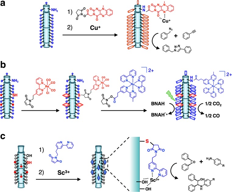
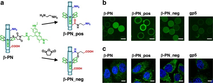
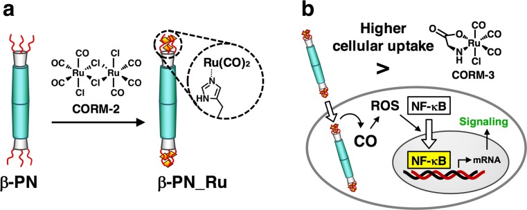
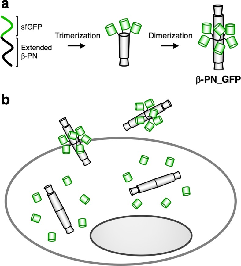
Bacteriophage T4 is a natural bio-nanomachine which achieves efficient infection of host cells via cooperative motion of specific three-dimensional protein architectures. The relationships between the protein structures and their dynamic functions have recently been clarified. In this review we summarize the design principles for fabrication of nanomachines using the component proteins of bacteriophage T4 based on these recent advances. We focus on the protein needle known as gp5, which is located at the center of the baseplate at the end of the contractile tail of bacteriophage T4. This protein needle plays a critical role in directly puncturing host cells, and analysis has revealed that it contains a common motif used for cell puncture in other known injection systems, such as T6SS. Our artificial needle based on the β-helical domain of gp5 retains the ability to penetrate cells and can be engineered to deliver various cargos into living cells. Thus, the unique components of bacteriophage T4 and other natural nanomachines have great potential for use as molecular scaffolds in efforts to fabricate new bio-nanomachines.

Keywords: Bacteriophage T4; Cell penetration; Gp5; Protein needle; β-Helix.


Conflict of interest statement

Conflict of interest

Hiroshi Inaba declares that he has no conflict of interest. Takafumi Ueno declares that he has no conflict of interest.

Ethical approval

This article does not contain any studies with human participants or animals performed by the authors.

Figures

Fig. 1
Fig. 1
Bacteriophage T4 with detailed structures of the head, the packaging motor, the sheath, the tail fiber and the baseplate with the needle (adapted from Fokine et al. ; Aksyuk et al. ; Sun et al. ; Taylor et al. 2016)
Fig. 2
Fig. 2
Reported and proposed nanomaterials based on the component proteins of bacteriophage T4, by means of chemical and genetic engineering. a Incorporation of DNA and protein into the head and modifications of protein, peptide, fluorescent dye and metal nanoparticles on the surface of the head (Hou et al. ; Robertson et al. ; Tao et al. ; Liu et al. 2014). b A DNA transporter based on the packaging motor which is inserted into lipid bilayers. c Nano-assembled tubes consisting of sheath proteins (Daube et al. 2007). d Self-assembled fiber consisting of tail fiber proteins (Hyman et al. 2002). e Nano-architecture (Sugimoto et al. ; Ueno et al. 2006), biocatalyst (Yokoi et al. ; Yokoi et al. ; Inaba et al. 2012) and cell penetration and intracellular delivery (Inaba et al. , ; Sanghamitra et al. 2014) using a cell-puncturing needle
Fig. 3
Fig. 3
a Engineering of the head of bacteriophage T4 for delivery of genes and proteins into mammalian cells (adapted from Tao et al. 2013). b Template synthesis of metal nanoparticles on the head of bacteriophage T4 (upper) and transmission electronic microscopy (TEM) images of Fe/T4, Co/T4, and Ni/T4 structures (bottom) (adapted from Hou et al. 2010). HOC Highly antigenic outer-capsid protein, Soc small outer capsid protein, gp gene product, GFP green fluorescent protein, Pi inorganic phosphate
Fig. 4
Fig. 4
a The DNA packaging motor assembled at the portal vertex of the head of bacteriophage T4 (adapted from Kottadiel et al. 2012). Schematic is not to scale. b Single DNA molecule packaging using an optical tweezer (adapted from Fuller et al. 2007). Copyright (2007) National Academy of Sciences, Washington D.C.
Fig. 5
Fig. 5
Synthesis and assembly of gp18 polysheath (a) and gp15 hexameric rings (b) and co-assembly of gp18 and gp15 to form a nano-doughnut (c) (adapted from Daube et al. 2007). Scale bars of the TEM images: a 20 nm, b 10 nm, c 40 nm
Fig. 6
Fig. 6
Engineering of tail fibers of bacteriophage T4 (adapted from Hyman et al. 2002). a Electron micrograph of bacteriophage T4 with short tail fibers. The white line next to the tail fiber indicates the length of a wild-type tail fiber. b Concept for forming a protein fiber based on chimera proteins of gp36 and gp37. Copyright (2002) National Academy of Sciences, Washington D.C.
Fig. 7
Fig. 7
a Baseplate of bacteriophage T4 (Kostyuchenko et al. 2003), b gp5–gp27–gp5.4 (combined from PDB IDs: 1 K28 and 4KU0) (Kanamaru et al. 2002). Each color represents a different monomer
Fig. 8
Fig. 8
Side view (top) and bottom view (bottom) snapshots from the simulation of membrane penetration of gp5 (adapted from Nishima et al. 2011). The zone in light blue shows the initial position of the membrane
Fig. 9
Fig. 9
Protein needles from Myoviridae bacteriophages. a gp5 and gp5.4 from bacteriophage T4 (combined from PDB IDs: 1 K28 and 4KU0) (Kanamaru et al. 2002). b gpV from bacteriophage P2 (combined from PDB IDs: 3QR7 and 3QR8) (Browning et al. 2012). c gp138 from bacteriophage ϕ92 (PDB ID: 3PQI) (Browning et al. 2012). d gp45 from bacteriophage Mu (PDB ID: 3VTO) (Harada et al. 2013). Each color represents a different monomer. OB-fold domain Oligonucleotide/oligosaccharide-binding domain
Fig. 10
Fig. 10
T6SS, the bacterial type VI secretion system, as a nanomachine for molecular injection (adapted from Shneider et al. 2013). a Model organization of the T6SS baseplate. Molecules 1–5 indicate the predicted effector molecules. b The conserved domains consisting of the VgrG–PAAR complex in T6SS. c Crystal structure of a chimeric protein including gp5-VgrG and the PAAR complex (PDB ID: 4JIV)
Fig. 11
Fig. 11
Construction of the β-helical protein needle (β-PN) by genetic fusion of the β-helix of gp5 and foldon of fibritin from bacteriophage T4 (Yokoi et al. 2010)
Fig. 12
Fig. 12
a Schematic image and TEM images of a tetrapod assembly of gp5 via formation of an Au nanocluster (adapted from Ueno et al. 2006). b Schematic image and TEM image of the supramolecular assembly consisting of LisDps, a spherical cage protein, and gp5C, the C-terminal domain of gp5 (adapted from Sugimoto et al. 2006)
Fig. 13
Fig. 13
a Schematic drawing of construction of an Fe(III) protoporphyrin and gp27–gp5 composite to promote a catalytic sulfoxidation reaction (Koshiyama et al. 2008). b Schematic representation of hetero-modification of fluorescein (Fl, donor) and tetramethylrhodamine (TMR, acceptor) on gp27–gp5 to induce fluorescence resonance energy transfer (FRET) (Koshiyama et al. 2009)
Fig. 14
Fig. 14
Construction of biocatalysts based on β-PN. a Modification of flavins and subsequent Cu(I) coordination on β-PN for catalytic enhancement of an azide–alkyne [3 + 2] cycloaddition reaction (Yokoi et al. 2010). b Sequential modification of Re and Ru complexes on β-PN for photocatalytic reduction of CO2 to CO by electron transfer from the Ru to Re complex in the presence of BNAH (1-benzyl-1,4-dihydronicotinamide) under irradiation with visible light (Yokoi et al. 2011). c Semi-synthesis of a catalytic Sc(III) complex by coordination of the bpy (2,2′-bipyridyl) and the two –ROH groups of β-PN for epoxide ring-opening reactions (Inaba et al. 2012)
Fig. 15
Fig. 15
Cell penetration of β-PN (adapted from Sanghamitra et al. 2014). a Modifications of charged residues of ATTO520-labeled β-PN. b, c Confocal fluorescence images of uptake into human red blood cells (b) (scale bars 5 μm) and HeLa cells (c) (scale bars 10 μm). β-PN_pos, β-PN_neg Positively and negatively charged β-PN, respectively
Fig. 16
Fig. 16
β-PN as a carrier of carbon monoxide (CO) (adapted from Inaba et al. 2015a). a Construction of an Ru carbonyl–protein needle construct (β-PN_Ru). b Intracellular CO delivery by β-PN_Ru and the subsequent signaling pathway. CORM CO-releasing molecule, ROS reactive oxygen species, NF-κB nuclear factor-kappaB
Fig. 17
Fig. 17
β-PN as a carrier of proteins (adapted from Inaba et al. 2014b). a Synthesis of the superfolder green fluorescent protein (sfGFP)–protein needle construct β-PN_GFP. b Intracellular delivery of sfGFP by β-PN_GFP

Similar articles

Cited by

References

    1. Aksyuk AA, Leiman PG, Kurochkina LP, Shneider MM, Kostyuchenko VA, Mesyanzhinov VV, Rossmann MG. The tail sheath structure of bacteriophage T4: a molecular machine for infecting bacteria. EMBO J. 2009;28:821–829. doi: 10.1038/emboj.2009.36. - DOI - PMC - PubMed
    1. Aksyuk AA, Bowman VD, Kaufmann B, Fields C, Klose T, Holdaway HA, Fischetti VA, Rossmann MG. Structural investigations of a Podoviridae streptococcus phage C1, implications for the mechanism of viral entry. Proc Natl Acad Sci USA. 2012;109:14001–14006. doi: 10.1073/pnas.1207730109. - DOI - PMC - PubMed
    1. Archer MJ, Liu JL. Bacteriophage T4 nanoparticles as materials in sensor applications: variables that influence their organization and assembly on surfaces. Sensors. 2009;9:6298–6311. doi: 10.3390/s90806298. - DOI - PMC - PubMed
    1. Arisaka F, Yap ML, Kanamaru S, Rossmann MG. Molecular assembly and structure of the bacteriophage T4 tail. Biophys Rev. 2016;8:385–396. doi: 10.1007/s12551-016-0230-x. - DOI - PMC - PubMed
    1. Bartual SG, Otero JM, Garcia-Doval C, Llamas-Saiz AL, Kahn R, Fox GC, van Raaij MJ. Structure of the bacteriophage T4 long tail fiber receptor-binding tip. Proc Natl Acad Sci USA. 2010;107:20287–20292. doi: 10.1073/pnas.1011218107. - DOI - PMC - PubMed