. 2018 Apr;192(1):95-103.
doi: 10.1111/cei.13082. Epub 2017 Dec 11.

Soluble TAM receptor tyrosine kinases in rheumatoid arthritis: correlation with disease activity and bone destruction

Affiliations

Soluble TAM receptor tyrosine kinases in rheumatoid arthritis: correlation with disease activity and bone destruction

L Xu et al. Clin Exp Immunol. 2018 Apr.

Abstract

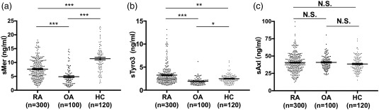
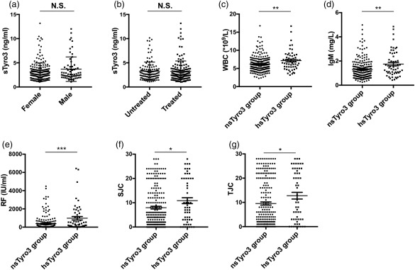
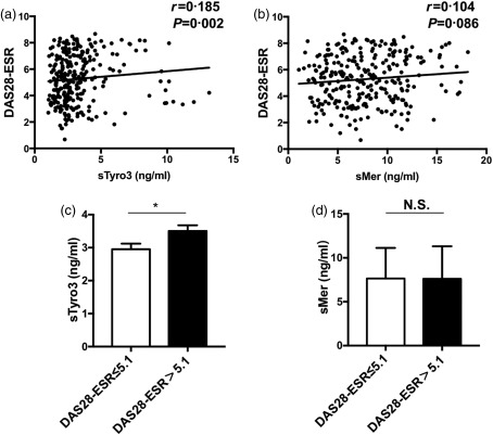
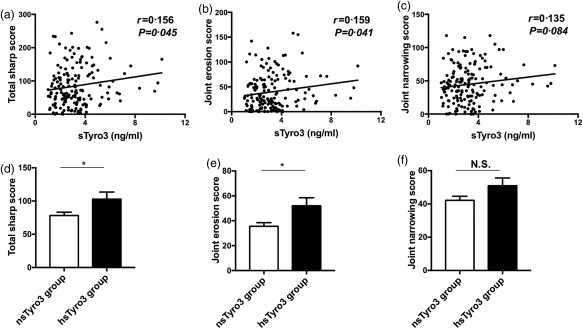
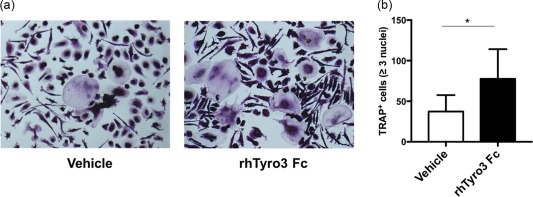
The TAM receptor tyrosine kinases (TAM RTK) are a subfamily of receptor tyrosine kinases, the role of which in autoimmune diseases such as systemic lupus erythematosus has been well explored, while their functions in rheumatoid arthritis (RA) remain largely unknown. In this study, we investigated the role of soluble TAM receptor tyrosine kinases (sAxl/sMer/sTyro3) in patients with RA. A total of 306 RA patients, 100 osteoarthritis (OA) patients and 120 healthy controls (HCs) were enrolled into this study. The serum concentrations of sAxl/sMer/sTyro3 were measured by enzyme-linked immunosorbent assay (ELISA), then the associations between sAxl/sMer/sTyro3 levels and clinical features of RA patients were analysed. We also investigated whether sTyro3 could promote osteoclast differentiation in vitro in RA patients. The results showed that compared with healthy controls (HCs), sTyro3 levels in the serum of RA patients were elevated remarkably and sMer levels were decreased significantly, whereas there was no difference between HCs and RA patients on sAxl levels. The sTyro3 levels were correlated weakly but positively with white blood cells (WBC), immunoglobulin (Ig)M, rheumatoid factor (RF), swollen joint counts, tender joint counts, total sharp scores and joint erosion scores. Conversely, there were no significant correlations between sMer levels and the above indices. Moreover, RA patients with high disease activity also showed higher sTyro3 levels. In-vitro osteoclast differentiation assay showed further that tartrate-resistant acid phosphatase (TRAP)+ osteoclasts were increased significantly in the presence of sTyro3. Collectively, our study indicated that serum sTyro3 levels were elevated in RA patients and correlated positively with disease activity and bone destruction, which may serve as an important participant in RA pathogenesis.

Keywords: TAM receptor tyrosine kinases; bone destruction; disease activity; rheumatoid arthritis.


Figures

Figure 1
Figure 1
Soluble Mer (sMer) (a), soluble Tyro3 (sTyro3) (b) and soluble Axl (sAxl) (c) levels in sera of rheumatoid arthritis (RA) patients, osteoarthritis (OA) patients and healthy controls (HC). (a) The concentrations of sMer were decreased in RA patients and OA patients compared with healthy controls. (b) sTyro3 levels were significantly increased relative to OA patients and healthy controls. (c) sAxl levels had no difference among RA patients, OA patients and healthy controls. The analyses were performed by covariance analysis (Dunnett's T3 test for post‐hoc test), *P < 0·05; **P < 0·01; ***P < 0·001; N.S. = not significant.
Figure 2
Figure 2
Associations between soluble Tyro3 (sTyro3) levels and clinical features of rheumatoid arthritis (RA) patients. Patients were divided by 90% confidence interval (1·0–4·0 ng/ml) from healthy controls for sTyro3 levels calculated by spss version 19.0: patients with sTyro3 levels greater than 4·0 ng/ml were grouped into the high sTyro3 (hsTyro3) group; sTyro3 levels of all other patients were between 1·0 and 4·0 ng/ml and these patients were grouped into the normal sTyro3 (nsTyro3) group. No difference was found between female and male patients (a), treated and untreated patients (b) on sTyro3 levels. White blood cells (WBC) (c), immunoglobulin (Ig)M (d), rheumatoid factor (RF) (e), swollen joint counts (SJC) (f) and tender joint counts (TJC) (g) were increased significantly in the hsTyro3 group. The analyses were performed by Mann–Whitney U test, *P < 0·05; **P < 0·01, ***P < 0·001.
Figure 3
Figure 3
Associations between soluble Mer and Tyro3 levels and rheumatoid arthritis (RA) patient disease activity score based on erythrocyte sedimentation rate (ESR) [28‐joint disease activity score (DAS28)‐ESR]. The correlation analyses between soluble Mer (sMer) / soluble Tyro3 (sTyro3) levels and DAS28‐ESR were performed by Spearman's correlation coefficient (a, b). sTyro3 levels in patients with DAS28‐ESR > 5·1 were higher than those of patients with DAS28‐ESR scores ≤ 5·1 (c), while no difference was indicated between two groups on sMer levels (d). The analyses were performed by Mann–Whitney U‐test, *P < 0·05; N.S. = not significant.
Figure 4
Figure 4
Correlations between soluble Tyro3 (sTyro3) levels and rheumatoid arthritis (RA) patient total Sharp scores, joint space narrowing scores and erosion scores. Positive correlations between sTyro3 levels and total Sharp scores (a), joint erosion scores (b) were found, but no correlation was found between sTyro3 levels and joint space narrowing scores (c). Next, we also grouped these patients as mentioned in Fig. 2; significant difference was shown between the high (hsTyro3) and normal sTyro3 (nsTyro3) groups with total Sharp scores (d) and joint erosion scores (e), but not joint space narrowing scores (f). The analyses were performed by Spearman's correlation coefficient and Mann–Whitney U test, *P < 0·05, N.S. = not significant.
Figure 5
Figure 5
Soluble Tyro3 (sTyro3) promoted osteoclast differentiation in rheumatoid arthritis (RA) patients. Freshly isolated peripheral blood mononuclear cells (PBMCs) were cultured with or without rhTyro3 Fc (500 ng/ml) under the stimulation of recombinant human macrophage colony‐stimulating factor (rhM‐CSF) (30 ng/ml) and recombinant human receptor activator of nuclear factor kappa‐Β ligand (rhRANKL) (50 ng/ml). Medium was changed every 3 days. Then, cells were subjected to tartrate‐resistant acid phosphatase (TRAP) staining on day 17. One representative experiment of six (a) and the statistical results were shown (b). The analysis was performed by Wilcoxon's matched‐pairs signed‐rank test, *P < 0·05. [Colour figure can be viewed at wileyonlinelibrary.com]

References

    1. Rothlin CV, Carrera‐Silva EA, Bosurgi L, Ghosh S. TAM receptor signaling in immune homeostasis. Annu Rev Immunol 2015; 33:355–91. - PMC - PubMed
    1. Lemke G, Rothlin CV. Immunobiology of the TAM receptors. Nat Rev Immunol 2008; 8:327–36. - PMC - PubMed
    1. Seitz HM, Camenisch TD, Lemke G, Earp HS, Matsushima GK. Macrophages and dendritic cells use different Axl/Mertk/Tyro3 receptors in clearance of apoptotic cells. J Immunol 2007; 178:5635–42. - PubMed
    1. Scutera S, Fraone T, Musso T et al Survival and migration of human dendritic cells are regulated by an IFN‐alpha‐inducible Axl/Gas6 pathway. J Immunol 2009; 183:3004–13. - PubMed
    1. Hilliard BA, Zizzo G, Ulas M, Linan MK, Schreiter J, Cohen PL. Increased expression of Mer tyrosine kinase in circulating dendritic cells and monocytes of lupus patients: correlations with plasma interferon activity and steroid therapy. Arthritis Res Ther 2014; 16:R76. - PMC - PubMed

Publication types

MeSH terms