

Resting-state fMRI in sleeping infants more closely resembles adult sleep than adult wakefulness
- PMID: 29149191
- PMCID: PMC5693436
- DOI: 10.1371/journal.pone.0188122
Resting-state fMRI in sleeping infants more closely resembles adult sleep than adult wakefulness
Abstract
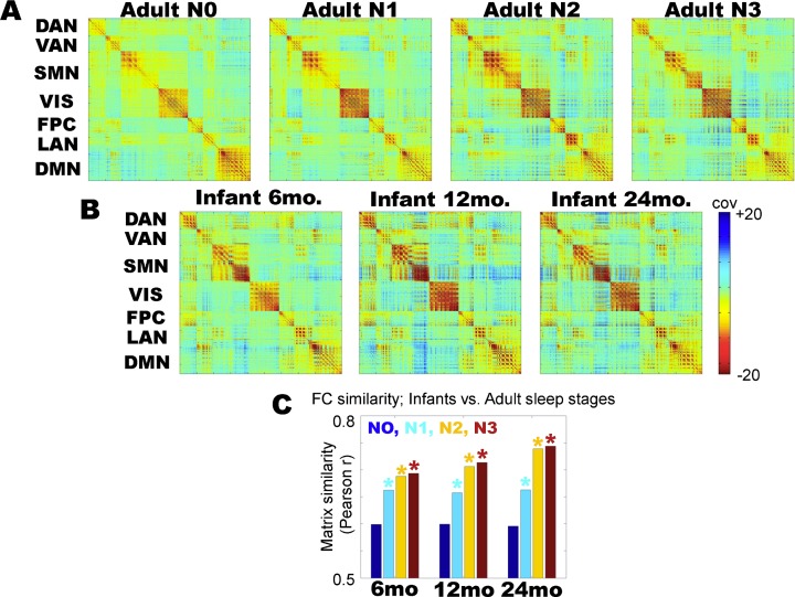
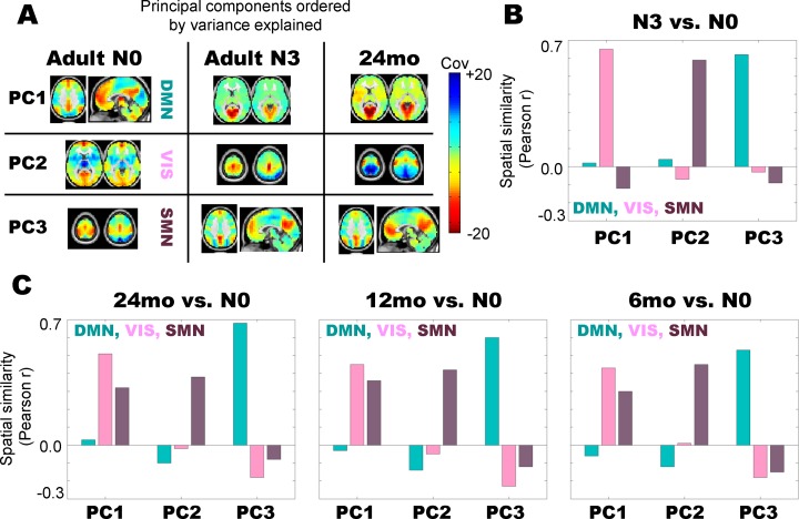
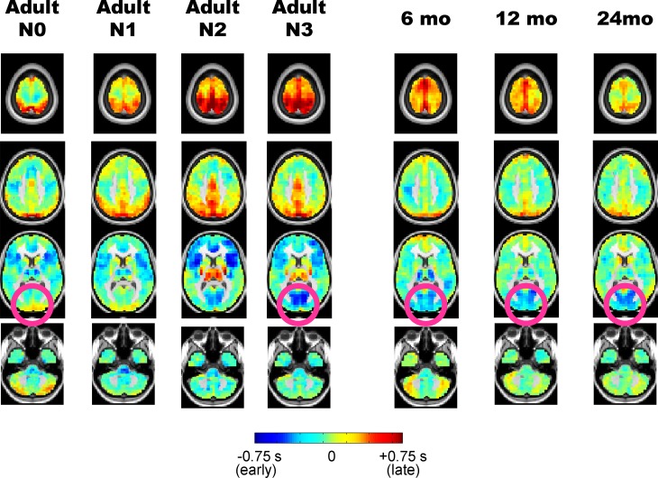
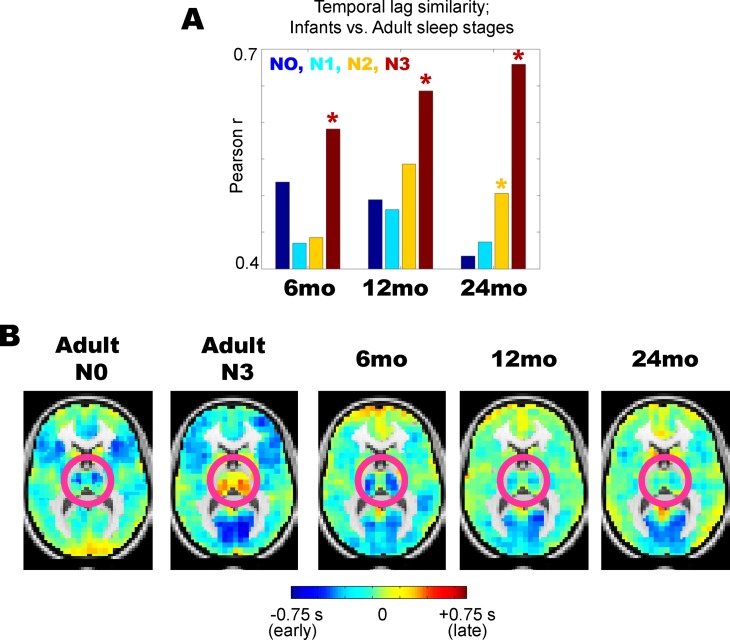
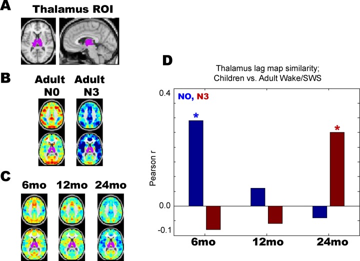
Resting state functional magnetic resonance imaging (rs-fMRI) in infants enables important studies of functional brain organization early in human development. However, rs-fMRI in infants has universally been obtained during sleep to reduce participant motion artifact, raising the question of whether differences in functional organization between awake adults and sleeping infants that are commonly attributed to development may instead derive, at least in part, from sleep. This question is especially important as rs-fMRI differences in adult wake vs. sleep are well documented. To investigate this question, we compared functional connectivity and BOLD signal propagation patterns in 6, 12, and 24 month old sleeping infants with patterns in adult wakefulness and non-REM sleep. We find that important functional connectivity features seen during infant sleep closely resemble those seen during adult sleep, including reduced default mode network functional connectivity. However, we also find differences between infant and adult sleep, especially in thalamic BOLD signal propagation patterns. These findings highlight the importance of considering sleep state when drawing developmental inferences in infant rs-fMRI.
Conflict of interest statement
Figures
Similar articles
-
Toward a complete taxonomy of resting state networks across wakefulness and sleep: an assessment of spatially distinct resting state networks using independent component analysis.Sleep. 2019 Mar 1;42(3):zsy235. doi: 10.1093/sleep/zsy235. Sleep. 2019. PMID: 30476346
-
Hard to wake up? The cerebral correlates of sleep inertia assessed using combined behavioral, EEG and fMRI measures.Neuroimage. 2019 Jan 1;184:266-278. doi: 10.1016/j.neuroimage.2018.09.033. Epub 2018 Sep 14. Neuroimage. 2019. PMID: 30223060
-
Resisting Sleep Pressure: Impact on Resting State Functional Network Connectivity.Brain Topogr. 2017 Nov;30(6):757-773. doi: 10.1007/s10548-017-0575-x. Epub 2017 Jul 15. Brain Topogr. 2017. PMID: 28712063
-
Resting-state functional MRI studies on infant brains: A decade of gap-filling efforts.Neuroimage. 2019 Jan 15;185:664-684. doi: 10.1016/j.neuroimage.2018.07.004. Epub 2018 Jul 7. Neuroimage. 2019. PMID: 29990581 Free PMC article. Review.
-
Spontaneous fMRI activity during resting wakefulness and sleep.Prog Brain Res. 2011;193:295-305. doi: 10.1016/B978-0-444-53839-0.00019-3. Prog Brain Res. 2011. PMID: 21854970 Free PMC article. Review.
Cited by
-
Hippocampal functional connectivity development during the first two years indexes 4-year working memory performance.Cortex. 2021 May;138:165-177. doi: 10.1016/j.cortex.2021.02.005. Epub 2021 Feb 17. Cortex. 2021. PMID: 33691225 Free PMC article.
-
Baby Brains at Work: How Task-Based Functional Magnetic Resonance Imaging Can Illuminate the Early Emergence of Psychiatric Risk.Biol Psychiatry. 2023 May 15;93(10):880-892. doi: 10.1016/j.biopsych.2023.01.010. Epub 2023 Jan 20. Biol Psychiatry. 2023. PMID: 36935330 Free PMC article. Review.
-
Machine Learning and Prediction in Fetal, Infant, and Toddler Neuroimaging: A Review and Primer.Biol Psychiatry. 2023 May 15;93(10):893-904. doi: 10.1016/j.biopsych.2022.10.014. Epub 2022 Oct 29. Biol Psychiatry. 2023. PMID: 36759257 Free PMC article.
-
Music in premature infants enhances high-level cognitive brain networks.Proc Natl Acad Sci U S A. 2019 Jun 11;116(24):12103-12108. doi: 10.1073/pnas.1817536116. Epub 2019 May 28. Proc Natl Acad Sci U S A. 2019. PMID: 31138687 Free PMC article.
-
Imaging the rapidly developing brain: Current challenges for MRI studies in the first five years of life.Dev Cogn Neurosci. 2021 Feb;47:100893. doi: 10.1016/j.dcn.2020.100893. Epub 2020 Dec 11. Dev Cogn Neurosci. 2021. PMID: 33341534 Free PMC article. Review.
References
-
- Lin W, Zhu Q, Gao W, Chen Y, Toh CH, Styner M, et al. Functional connectivity MR imaging reveals cortical functional connectivity in the developing brain. AJNR Am J Neuroradiol. 2008;29(10):1883–9. doi: 10.3174/ajnr.A1256 ; PubMed Central PMCID: PMC2583167. - DOI - PMC - PubMed
-
- Fransson P, Skiold B, Engstrom M, Hallberg B, Mosskin M, Aden U, et al. Spontaneous brain activity in the newborn brain during natural sleep—an fMRI study in infants born at full term. Pediatr Res. 2009;66(3):301–5. doi: 10.1203/PDR.0b013e3181b1bd84 . - DOI - PubMed
-
- Pruett JR Jr., Kandala S, Hoertel S, Snyder AZ, Elison JT, Nishino T, et al. Accurate age classification of 6 and 12 month-old infants based on resting-state functional connectivity magnetic resonance imaging data. Dev Cogn Neurosci. 2015;12:123–33. doi: 10.1016/j.dcn.2015.01.003 ; PubMed Central PMCID: PMC4385423. - DOI - PMC - PubMed
-
- Gao W, Zhu H, Giovanello KS, Smith JK, Shen D, Gilmore JH, et al. Evidence on the emergence of the brain's default network from 2-week-old to 2-year-old healthy pediatric subjects. Proc Natl Acad Sci U S A. 2009;106(16):6790–5. doi: 10.1073/pnas.0811221106 ; PubMed Central PMCID: PMC2672537. - DOI - PMC - PubMed
-
- Smyser CD, Snyder AZ, Shimony JS, Mitra A, Inder TE, Neil JJ. Resting-State Network Complexity and Magnitude Are Reduced in Prematurely Born Infants. Cereb Cortex. 2014. doi: 10.1093/cercor/bhu251 . - DOI - PMC - PubMed
MeSH terms
Grants and funding
LinkOut - more resources
Full Text Sources
Other Literature Sources
