Mycolactone displays anti-inflammatory effects on the nervous system
- PMID: 29149212
- PMCID: PMC5693295
- DOI: 10.1371/journal.pntd.0006058
Mycolactone displays anti-inflammatory effects on the nervous system
Abstract
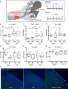
Background: Mycolactone is a macrolide produced by the skin pathogen Mycobacterium ulcerans, with cytotoxic, analgesic and immunomodulatory properties. The latter were recently shown to result from mycolactone blocking the Sec61-dependent production of pro-inflammatory mediators by immune cells. Here we investigated whether mycolactone similarly affects the inflammatory responses of the nervous cell subsets involved in pain perception, transmission and maintenance. We also investigated the effects of mycolactone on the neuroinflammation that is associated with chronic pain in vivo.
Methodology/ principle findings: Sensory neurons, Schwann cells and microglia were isolated from mice for ex vivo assessment of mycolactone cytotoxicity and immunomodulatory activity by measuring the production of proalgesic cytokines and chemokines. In all cell types studied, prolonged (>48h) exposure to mycolactone induced significant cell death at concentrations >10 ng/ml. Within the first 24h treatment, nanomolar concentrations of mycolactone efficiently suppressed the cell production of pro-inflammatory mediators, without affecting their viability. Notably, mycolactone also prevented the pro-inflammatory polarization of cortical microglia. Since these cells critically contribute to neuroinflammation, we next tested if mycolactone impacts this pathogenic process in vivo. We used a rat model of neuropathic pain induced by chronic constriction of the sciatic nerve. Here, mycolactone was injected daily for 3 days in the spinal canal, to ensure its proper delivery to spinal cord. While this treatment failed to prevent injury-induced neuroinflammation, it decreased significantly the local production of inflammatory cytokines without inducing detectable cytotoxicity.
Conclusion/ significance: The present study provides in vitro and in vivo evidence that mycolactone suppresses the inflammatory responses of sensory neurons, Schwann cells and microglia, without affecting the cell viability. Together with previous studies using peripheral blood leukocytes, our work implies that mycolactone-mediated analgesia may, at least partially, be explained by its anti-inflammatory properties.
Conflict of interest statement
The authors have declared that no competing interests exist.
Figures


Similar articles
-
Antibody-Mediated Neutralization of the Exotoxin Mycolactone, the Main Virulence Factor Produced by Mycobacterium ulcerans.PLoS Negl Trop Dis. 2016 Jun 28;10(6):e0004808. doi: 10.1371/journal.pntd.0004808. eCollection 2016 Jun. PLoS Negl Trop Dis. 2016. PMID: 27351976 Free PMC article.
-
Mycobacterium ulcerans toxic macrolide, mycolactone modulates the host immune response and cellular location of M. ulcerans in vitro and in vivo.Cell Microbiol. 2005 Sep;7(9):1295-304. doi: 10.1111/j.1462-5822.2005.00557.x. Cell Microbiol. 2005. PMID: 16098217
-
Shaping mycolactone for therapeutic use against inflammatory disorders.Sci Transl Med. 2015 May 27;7(289):289ra85. doi: 10.1126/scitranslmed.aab0458. Sci Transl Med. 2015. PMID: 26019221
-
Immunity against Mycobacterium ulcerans: The subversive role of mycolactone.Immunol Rev. 2021 May;301(1):209-221. doi: 10.1111/imr.12956. Epub 2021 Feb 19. Immunol Rev. 2021. PMID: 33607704 Review.
-
Pleiotropic molecular effects of the Mycobacterium ulcerans virulence factor mycolactone underlying the cell death and immunosuppression seen in Buruli ulcer.Biochem Soc Trans. 2014 Feb;42(1):177-83. doi: 10.1042/BST20130133. Biochem Soc Trans. 2014. PMID: 24450648 Review.
Cited by
-
The One That Got Away: How Macrophage-Derived IL-1β Escapes the Mycolactone-Dependent Sec61 Blockade in Buruli Ulcer.Front Immunol. 2022 Jan 26;12:788146. doi: 10.3389/fimmu.2021.788146. eCollection 2021. Front Immunol. 2022. PMID: 35154073 Free PMC article.
-
Neuronal Regulation of Immunity in the Skin and Lungs.Trends Neurosci. 2019 Aug;42(8):537-551. doi: 10.1016/j.tins.2019.05.005. Epub 2019 Jun 15. Trends Neurosci. 2019. PMID: 31213389 Free PMC article. Review.
-
Could Mycolactone Inspire New Potent Analgesics? Perspectives and Pitfalls.Toxins (Basel). 2019 Sep 4;11(9):516. doi: 10.3390/toxins11090516. Toxins (Basel). 2019. PMID: 31487908 Free PMC article. Review.
-
Mycolactone as Analgesic: Subcutaneous Bioavailability Parameters.Front Pharmacol. 2019 Apr 12;10:378. doi: 10.3389/fphar.2019.00378. eCollection 2019. Front Pharmacol. 2019. PMID: 31031626 Free PMC article.
-
Aberrant stromal tissue factor localisation and mycolactone-driven vascular dysfunction, exacerbated by IL-1β, are linked to fibrin formation in Buruli ulcer lesions.PLoS Pathog. 2022 Jan 31;18(1):e1010280. doi: 10.1371/journal.ppat.1010280. eCollection 2022 Jan. PLoS Pathog. 2022. PMID: 35100311 Free PMC article. Clinical Trial.
References
-
- George KM, Chatterjee D, Gunawardana G, Welty D, Hayman J, Lee R, et al. Mycolactone: a polyketide toxin from Mycobacterium ulcerans required for virulence. Science. 1999;283(5403):854–7. . - PubMed
-
- Demangel C, Stinear TP, Cole ST. Buruli ulcer: reductive evolution enhances pathogenicity of Mycobacterium ulcerans. Nat Rev Microbiol. 2009;7(1):50–60. doi: 10.1038/nrmicro2077 . - DOI - PubMed
-
- Hall B, Simmonds R. Pleiotropic molecular effects of the Mycobacterium ulcerans virulence factor mycolactone underlying the cell death and immunosuppression seen in Buruli ulcer. Biochem Soc Trans. 2014;42(1):177–83. doi: 10.1042/BST20130133 . - DOI - PubMed
-
- Guenin-Mace L, Baron L, Chany AC, Tresse C, Saint-Auret S, Jonsson F, et al. Shaping mycolactone for therapeutic use against inflammatory disorders. Sci Transl Med. 2015;7(289):289ra85 doi: 10.1126/scitranslmed.aab0458 . - DOI - PubMed
-
- Zavattaro E, Boccafoschi F, Borgogna C, Conca A, Johnson RC, Sopoh GE, et al. Apoptosis in Buruli ulcer: a clinicopathological study of 45 cases. Histopathology. 2012;61(2):224–36. doi: 10.1111/j.1365-2559.2012.04206.x . - DOI - PubMed
MeSH terms
Substances
LinkOut - more resources
Full Text Sources
Other Literature Sources

