. 2017 Nov 16;68(4):673-685.e6.
doi: 10.1016/j.molcel.2017.10.022.

Sterol Oxidation Mediates Stress-Responsive Vms1 Translocation to Mitochondria

Affiliations

Sterol Oxidation Mediates Stress-Responsive Vms1 Translocation to Mitochondria

Jason R Nielson et al. Mol Cell. .

Abstract

Vms1 translocates to damaged mitochondria in response to stress, whereupon its binding partner, Cdc48, contributes to mitochondrial protein homeostasis. Mitochondrial targeting of Vms1 is mediated by its conserved mitochondrial targeting domain (MTD), which, in unstressed conditions, is inhibited by intramolecular binding to the Vms1 leucine-rich sequence (LRS). Here, we report a 2.7 Å crystal structure of Vms1 that reveals that the LRS lies in a hydrophobic groove in the autoinhibited MTD. We also demonstrate that the oxidized sterol, ergosterol peroxide, is necessary and sufficient for Vms1 localization to mitochondria, through binding the MTD in an interaction that is competitive with binding to the LRS. These data support a model in which stressed mitochondria generate an oxidized sterol receptor that recruits Vms1 to support mitochondrial protein homeostasis.

Keywords: ROS signaling; S. cerevisiae; biochemistry; lipid signaling; liposomes; mitochondrial quality control; oxidative stress; protein degradation; sterols; structure-function.


Figures

Figure 1
Figure 1. Structure of Vms1 region that regulates mitochondrial localization
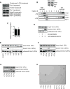
(A) Schematic representation of the domain structure of Vms1. Black line below indicates the crystallized construct. LRS, Leucine Rich Sequence; ZnF, Zinc Finger; MTD, Mitochondrial Targeting Domain; AnkR, Ankryin Repeat; CC, Coil-Coil; VIM, VCP-Interacting Motif. Greyed out areas, including all segments between domains, denotes regions not visible in the electron density maps. (B) Vms1LRS-ZnF-MTD ribbon representation. The Zn ion is represented by a gray sphere. Dashed lines indicate residues not visible in the structure, except for the large segments 36-73 and 108-188, the ends of which are labeled with residue numbers. (C) Alignment of LRS residues visible in (B). White letters with gray background indicates similarity. White letters with a black background indicates identity. Yellow asterisks indicate Leu residues whose mutation abolished MTD interaction (Heo et al, 2013). (D) LRS Leu 23, 28, 31, and 33 (yellow), the same residues marked by asterisks in (C), are shown explicitly on the Vms1 ribbon representation. The MTD is shown as a surface representation, colored by K-D hydrophobicity (Kyte and Doolittle, 1982). (E) Alignment of MTD hydrophobic groove that contacts the LRS. Conservation is colored as in (C). Blue asterisks indicate residues shown in (F). (F) Ribbon representation showing the interaction between the MTD and LRS on the left. Side chains are shown for hydrophobic residues in each domain that are buried at the LRS-MTD interface. The LRS is removed on the right to better visualize the MTD residues. See also Figure S1.
Figure 2
Figure 2. MTD surface surrounding the Vms1LRS-MTD interface mediates localization to mitochondria
(A) vms1Δ cells expressing the indicated Vms1MTD-GFP construct and mitochondria-targeted RFP were grown to mid-log phase and analyzed by fluorescence microscopy. Representative images are shown. (B) The mitochondrial MTD-GFP intensity from (A) was quantified as described for 100+ cells in each strain over multiple days of imaging. (C) Cells from (A) were lysed as described and analyzed by Western blot. (D) Table of MTD residues mutated, with quantified mitochondrial localization relative to WT (+/− SEM) and p value indicated. Residues were initially mutated in pairs or triplets, and only made as single mutants if there was a greater than 10% reduction in MTD localization. Some mutants enhanced mitochondrial localization, indicated by values greater than one. The far-right column gives the percent increase in relative Å2 surface exposure of each amino acid in the MTD alone vs the LRS-MTD complex, based on assuming no conformational change upon removing the LRS from the crystal structure. (E) Ribbon/surface overview of Vms1MTD with mutated residues highlighted. Residues whose mutation exhibited >10% reduction (red) and ≤10% reduction (green) in MTD localization are indicated. (F) The vms1Δ strain was transformed with Vms11-182-HA and the indicated Vms1MTD-GFP construct. Strain lysates were immunoprecipitated with anti-HA antibody. Western blots were performed with anti-HA and GFP antibodies.
Figure 3
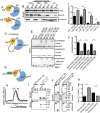
Figure 3. Vms1 binds a lipid with molecular formula C28H44O3
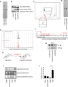
(A) Purified mitochondria treated with or without proteinase K were subjected to western blot (20μg loaded). (B) Purified Vms1 and untreated or proteinase K-treated mitochondria were mixed and subjected to sucrose gradient centrifugation. Fractions were collected and subjected to Western blot. (C) Co-migration of Vms1 and Porin from (B) was quantitated over at least 5 experiments in each condition (mean ± SEM). (D) Floatation assay results for control liposomes and liposomes supplemented with mitochondrial lipids. For liposomes containing mitochondrial lipids, we added lipids isolated from 1mg of mitochondria (determined by measuring protein concentration) per 100μL liposomes. Purified mitochondrial lipids were added either without modification or following akaline-treatment to generate alkaline-resistant mitochondrial lipids. (E) Floatation assay results obtained with liposomes prepared with UPLC fractions of purified mitochondrial lipids (lipids from 2mg mitochondria per 100μL liposomes). (F) Fraction 2 in (E) was sub-fractionated and resulting lipid fractions were incorporated into liposomes (lipids from 1mg mitochondria per 100μL liposomes) and subjected to a floatation assay. (G) Mitochondrial alkaline-resistant lipids were separated into 5 fractions by TLC. Lipids isolated from each fraction were incorporated into liposomes (lipids from 1mg mitochondria per 100μL liposomes) and subjected to a floatation assay. (H) Lipids from the 5 UPLC sub-fractions (F) and the 5 TLC fractions (G) were analyzed by TLC and orcinol stain. The red star indicates the common species with formula C28H44O3.
Figure 4
Figure 4. The C28H44O3 lipid is ergosterol peroxide, which binds Vms1 directly
(A) The C28H44O3 species (red star) was isolated from mitochondrial alkaline-resistant lipids by silica chromatography. (B) Liposomes containing the silica chromatography input or the isolated C28H44O3 species were prepared such that they had equivalent amounts of C28H44O3 (lipids from approximately 1mg mitochondria per 100μL liposomes) and subjected to a floatation assay. (C) 1-H NMR spectrum of the isolated C28H44O3. (D) Comparison of the isolated C28H44O3 species and commercial ergosterol peroxide by TLC and orcinol stain. (E) Comparison of the isolated C28H44O3 species and commercial ergosterol peroxide by LC/MS. (F) Comparison of the isolated C28H44O3 species (red) and commercial ergosterol peroxide (black) by MS/MS at 20 eV. (G) Nitrocellulose membranes were spotted with equivalent amounts (5 μg-top and 1.67 μg-bottom) of isolated C28H44O3 species, ergosterol peroxide, or ergosterol and assayed for binding to Vms1 by a modified immunoblot assay. (H) The non-enzymatic oxidation of ergosterol to ergosterol peroxide. (I) WT cells were fractionated into whole cell extract (WCE), spheroplasts, post-mitochondrial supernatant (PMS), and mitochondria. Lipids from 2.5 mg of each fraction (by protein concentration) were analyzed by TLC and orcinol/primuline stain. (J) Ergosterol peroxide from whole cell extract (WCE), extract after cell wall removal (Spheroplast), post-mitochondrial supernatant (PMS), and crude mitochondria (Mito) was quantified as the ratio of ergosterol peroxide to protein concentration over at least 3 experiments. **p≤0.01. *p≤0.05. See also Figure S2.
Figure 5
Figure 5. Ergosterol peroxide is necessary for Vms1 localization to mitochondria
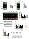
(A) WT cells expressing Vms1MTD-GFP and mitochondria-targeted RFP were grown for 6 hours in anoxia or normoxia and subjected to fluorescence microscopy. Representative images are shown. (B) The mitochondrial MTD intensity was quantified as described for 100+ cells in each condition in (A). ***p≤0.001. (C) Cells from (A) were grown in the presence of 500 μM mevastatin or vehicle for 24 hours and subjected to fluorescence microscopy. Representative images are shown. (D) The mitochondrial MTD intensity was quantified as described for 100+ cells in each condition in (C). ***p≤0.001. (E) WT and the indicated mutants expressing Vms1MTD-GFP and mitochondria-targeted RFP were grown to mid-log phase and subjected to fluorescence microscopy. Representative images are shown. (F) The mitochondrial MTD intensity was quantified as described for 50+ cells in each strain in (E). ***p≤0.001. **p≤0.01. *p≤0.05. (G) 20 μg of purified mitochondria (by protein concentration) from the indicated strains were analyzed by Western blot as a loading control (top). Lipids from 2 mg of mitochondria from each strain were analyzed by TLC and orcinol stain. (H) Ergosterol peroxide* from (G) was quantitated as the ratio of ergosterol peroxide* to Porin over 3 independent experiments. ***p≤0.001. *p≤0.05. (I) Structure of ergosterol indicating bonds and enzymes whose loss impairs Vms1 localization to mitochondria (red) and those that leave localization unaffected (blue). (J) Vehicle, 3.75 μg ergosterol, or 3.75 μg ergosterol peroxide were added to mitochondrial lipids isolated from 300 μg of WT or erg2Δ cells to make liposomes as described (See Methods) for use in the floatation assay. (K) Vms1 binding to liposomes from (J) was quantified as the ratio of bound Vms1 to input Vms1 and normalized to liposomes containing no added lipids for each strain. **p≤0.01. See also Figure S3.
Figure 6
Figure 6. Increased ergosterol peroxide levels in response to stress correlate with increased Vms1 binding to mitochondrial lipids
(A) Purified Vms1 and mitochondria isolated from vehicle, rapamycin (rapa), or H2O2-treated cells were co-incubated and subjected to sucrose gradient centrifugation. (B) Co-migration was quantitated from (A) as the ratio of Vms1 to Porin over at least 4 independent experiments. **p≤0.01. *p≤0.05. (C) 20 μg of purified mitochondria (by protein concentration) from the indicated treatments were analyzed by Western blot. Lipids from 2 mg of mitochondria from each strain were analyzed by TLC and orcinol stain. (D) Ergosterol peroxide from (D) was quantitated as the ratio of ergosterol peroxide to Porin over at least 3 experiments. ***p≤0.001. *p≤0.05. (E) 100 μg of mitochondrial lipids isolated from cells subjected to the indicated treatments were used to make liposomes for floatation assays. (F) Vms1 binding from (F) was quantified as the ratio of bound Vms1 to input over at least 4 experiments. *p≤0.05.
Figure 7
Figure 7. LRS and lipid compete for MTD binding
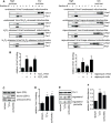
(A) WT and L4A representations indicating that Leu to Ala mutations in L4A weaken the interaction with the MTD. (B) Representative floatation assay of Vms1WT or Vms1L4A following incubation with liposomes (3, 10, 30, 100 μL) containing 100 μg of erg2Δ lipids with 1.25 μg of exogenous EP per 100 μL. (C) Quantification of at least 3 floatation assay experiments from (B). Vms1 binding to liposomes was quantified as a ratio of the bound/input. **p≤0.01. *p≤0.05. (D) Cysteine residues were introduced into the Vms1LRS and proximally in the Vms1MTD. A PreScission protease (pp) site was introduced between the two domains to allow for observation of crosslinking efficiency on SDS-PAGE. (E) Representative floatation assay of the indicated WT or mutant His10-Vms11-417-HA constructs. Proteins with and without BMH crosslinking following incubation with PreScission protease were incubated with liposomes consisting of base lipids (DOPC, ceramide-OH, ergosterol) and 1.25 μg EP. The protein/liposomes mixture was then subjected to the floatation assay. (F) Crosslinked/cleaved protein from (E) was quantified in the input and bound fractions. **p≤0.01. (G) His12-GFP-Vms1LRS-pp-Vms1MTD-HA construct allows tracking of GFP-Vms1LRS and Vms1MTD-HA liposome binding within a single experiment. (H) Size exclusion chromatogram of GFP-Vms1LRS-pp-Vms1MTD-HA shows equivalent elution with or without cleavage by PreScission protease. Sizing standard peaks are indicated by dashed lines. (I) Western blots of fractions (1-3) from (H) shows equivalent elution with or without cleavage by PreScission protease. (J) Representative floatation assay of cleaved (GFP-Vms1LRS/Vms1MTD-HA) and uncleaved proteins (GFP-Vms1full-HA) from (H) incubated with liposomes containing 100 μg of erg2Δ lipids with 1.25 μg of exogenous EP. (K) Vms1full/Vms1cleaved ratio from experiment in (H) was quantified in the bound and unbound fractions for both GFP and HA. **p≤0.01. See also Figure S4.

References

    1. Aberle D, Oetter KM, Meyers G. Lipid Binding of the Amphipathic Helix Serving as Membrane Anchor of Pestivirus Glycoprotein Erns. PloS one. 2015;10:e0135680. - PMC - PubMed
    1. Adams PD, Afonine PV, Bunkoczi G, Chen VB, Davis IW, Echols N, Headd JJ, Hung LW, Kapral GJ, Grosse-Kunstleve RW, et al. PHENIX: a comprehensive Python-based system for macromolecular structure solution. Acta crystallographica Section D, Biological crystallography. 2010;66:213–221. - PMC - PubMed
    1. Bagnat M, Keranen S, Shevchenko A, Shevchenko A, Simons K. Lipid rafts function in biosynthetic delivery of proteins to the cell surface in yeast. Proc Natl Acad Sci U S A. 2000;97:3254–3259. - PMC - PubMed
    1. Balaban RS, Nemoto S, Finkel T. Mitochondria, oxidants, and aging. Cell. 2005;120:483–495. - PubMed
    1. Bocking T, Barrow KD, Netting AG, Chilcott TC, Coster HG, Hofer M. Effects of singlet oxygen on membrane sterols in the yeast Saccharomyces cerevisiae. Eur J Biochem. 2000;267:1607–1618. - PubMed

MeSH terms