. 2018 Apr:14:576-587.
doi: 10.1016/j.redox.2017.11.004. Epub 2017 Nov 6.

DUSP1 alleviates cardiac ischemia/reperfusion injury by suppressing the Mff-required mitochondrial fission and Bnip3-related mitophagy via the JNK pathways

Affiliations

DUSP1 alleviates cardiac ischemia/reperfusion injury by suppressing the Mff-required mitochondrial fission and Bnip3-related mitophagy via the JNK pathways

Qinhua Jin et al. Redox Biol. 2018 Apr.

Abstract

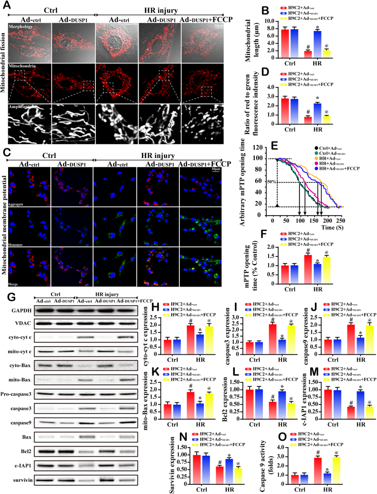
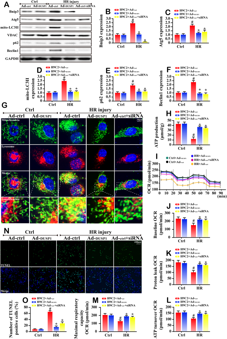
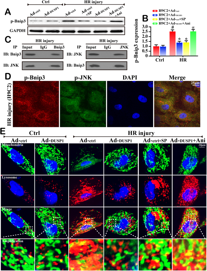
Mitochondrial fission and selective mitochondrial autophagy (mitophagy) form an essential axis of mitochondrial quality control that plays a critical role in the development of cardiac ischemia-reperfusion (IR) injury. However, the precise upstream molecular mechanism of fission/mitophagy remains unclear. Dual-specificity protein phosphatase1 (DUSP1) regulates cardiac metabolism, but its physiological contribution in the reperfused heart, particularly its influence on mitochondrial homeostasis, is unknown. Here, we demonstrated that cardiac DUSP1 was downregulated following acute cardiac IR injury. In vivo, compared to wild-type mice, DUSP1 transgenic mice (DUSP1TG mice) demonstrated a smaller infarcted area and the improved myocardial function. In vitro, the IR-induced DUSP1 deficiency promoted the activation of JNK which upregulated the expression of the mitochondrial fission factor (Mff). A higher expression level of Mff was associated with elevated mitochondrial fission and mitochondrial apoptosis. Additionally, the loss of DUSP1 also amplified the Bnip3 phosphorylated activation via JNK, leading to the activation of mitophagy. Increased mitophagy overtly consumed mitochondrial mass resulting into the mitochondrial metabolism disorder. However, the reintroduction of DUSP1 blunted Mff/Bnip3 activation and therefore alleviated the fatal mitochondrial fission/mitophagy by inactivating the JNK pathway, providing a survival advantage to myocardial tissue following IR stress. The results of our study suggest that DUSP1 and its downstream JNK pathway are therapeutic targets for conferring protection against IR injury by repressing Mff-mediated mitochondrial fission and Bnip3-required mitophagy.

Keywords: Bnip3; Cardiac IR injury; DUSP1; JNK; Mff; Mitochondrial fission; Mitophagy.


Figures

fx1
Graphical abstract
Fig. 1
Fig. 1
Downregulation of DUSP1 at the infarcted area. WT mice and DUSP1TG mice underwent the 2-h ischemia and 0–24-h reperfusion (IR injury, n = 6/group). In vitro, 2-h of hypoxia and 6-h of reoxygenation (HR) was used to mimic the IR injury. Meanwhile, the gain-of-function assay about DUSP1 was also conducted via adenovirus vector overexpression of DUSP1 (Ad-DUSP1) or control adenovirus transfection (Ad-ctrl) in H9C2 cells. A. DUSP1 expression in the infarcted area or non-infarcted area. B. Quantitative analysis of the relative expression of DUSP1. C-D. The immunohistochemistry of DUSP1 in heart underwent 2-h ischemia and 6-h reperfusion. E-F. Representative images of heart sections with TTC and Evans Blue staining of the infarcted area. Bar graph indicates the infarct size. G-H. TUNEL assay for cellular apoptosis analysis. I-J. The control adenovirus (Ad-ctrl) and DUSP1-overexpression adenovirus (Ad-DUSP1) were transfected into H9C2 cells. The transfection efficiency was confirmed by western blots. K-L. Caspase3 activity and MTT assay were used to detect the cellular viability and apoptosis. #P < 0.05 vs the sham group or control group; *P < 0.05 vs IR+WT group or HR+Ad-ctrl group.
Fig. 2
Fig. 2
DUSP1 influenced mitochondrial fission. A. Tom20 was used to observe the mitochondrial fission or mitochondrial debris. The bottom picture under each micrograph shows an amplification of mitochondrial fragments. B. The quantification of mitochondrial fission via mitochondrial length. FCCP, the fission activator, was used to re-activate fission in DUSP1-overexpressed cells. C-D. The change in the membrane potential (ΔΨm) via JC-1 staining. E-F. The mPTP opening time was recorded and the mPTP opening rate was evaluated with DUSP1 overexpression or not. G-N. Western blots was used to evaluate the change of mitochondrial apoptosis-related proteins. O. Caspase9 activity was measured to reflect the mitochondrial apoptosis. #P < 0.05 vs the control group; *P < 0.05 vs HR+Ad-ctrl group; @P < 0.05 vs HR+Ad-DUSP1 group.
Fig. 3
Fig. 3
DUSP1 handled fission via JNK/Mff pathways. A-B. The changes of mitochondrial fission receptors. Only Mff was increased in response to HR injury and was also handled by DUSP1. C-E. DUSP1 inactivated Mff via JNK. SP600125 (SP) is the inhibitor of JNK and it was used to block the HR-mediated JNK phosphorylation. Anisomycin (Ani) is the activator of JNK, which was used to re-activate the JNK activity in DUSP1-overepxressed cells. F. The co-localization of Drp1 and mitochondria. The pictures under each micrograph represent the amplification of the white square. More Drp1 was located on fragmented mitochondria, while the reintroduction of DUSP1 reduced the Drp1 migration on mitochondria. G. The qPCR assay was used to analyze the change of Mff expression. H-I. Chromatin-anti-p-JNK immunoprecipitation (ChIP) assay displayed that p-JNK interacted with the Mff promoter under HR injury, but it did not in DUSP1-overexpression or JNK inhibitor-treated cells. Immunoglobulin-G (IgG) was used as negative control, and anti-histone-3 (α-H3) as a positive control; input demonstrated equal amplification of DNA in non-immunoprecipitated samples; IP corresponds to the amplified DNA band in the immunoprecipitated samples. #P < 0.05 vs the control group; *P < 0.05 vs HR+Ad-ctrl group; @P < 0.05 vs HR+Ad-DUSP1 group.
Fig. 4
Fig. 4
DUSP1 overexpression was involved in mitophagy inhibition via Bnip3. A-F. Western blots was used to analyze the proteins related to mitophagy. The siRNA against Bnip3 was used to knockdown the expression of Bnip3, which was considered as the negative control group. G. The immunofluorescence assay of mitochondria and lysosome. In the amplified panel, the yellow fluorescence is indicative of mitophagy. H. The ATP production was measured with DUSP1 overepxression or Bnip3 silence. I-M. The OCR assay was used to obverse the mitochondrial respiratory function. Data are shown as the means ± SEM. O-N. TUNEL assay was used to explore the role of Bnip3-related mitophagy in cell death. #P < 0.05 vs the control group; *P < 0.05 vs HR+Ad-ctrl group.
Fig. 5
Fig. 5
DUSP1 inactivated Bnip3 via inhibiting JNK-mediated Bnip3 phosphorylation. A-B. Bnip3 phosphorylation was analyzed via western blots. SP600125 (SP) is the inhibitor of JNK and it was used to block the HR-mediated JNK phosphorylation. Anisomycin (Ani) is the activator of JNK, which was used to reactivate the JNK activity in DUSP1-overepxressed cells. C. Cell lysates were immunoprecipitated with anti-Bnip3 or anti-JNK antibodies, followed by immunoblotting with anti-JNK or anti-Bnip3 antibody. IgG was used as a control. D. The cytoplasmic localization of JNK-Bnip3 interaction has also been observed. The confocal microscopy showed approximately 80% colocalization of JNK and Bnip3 in the cytoplasm of H9C2 cells. E. The co-immunofluorescence of mitochondria and lysosomes. DUSP1 alleviated the overlap of mitochondria and lysosomes. #P < 0.05 vs the control group; *P < 0.05 vs HR+Ad-ctrl group; @P < 0.05 vs HR+Ad-DUSP1 group.
Fig. 6
Fig. 6
IR injury caused DUSP1 downregulation, leading to an increase in JNK phosphorylation. Activated JNK increased the expression of Mff, which initiated fatal mitochondrial fission. Excessive fission induced the pro-apoptotic cyt-c leakage from mitochondria into cytoplasm, launching the caspase9 related mitochondrial apoptosis. Meanwhile, JNK also phosphorylated Bnip3 and elevated mitophagy activity. Extensive mitophagy overtly consumed mitochondrial mass impairing the mitochondrial energy production. However, recovery of DUSP1 activity could cut off the excessive fission and mitophagy, providing pro-survival advantages for the heart in the context of IR injury.
Fig. S1
Fig. S1
Reintroduction of DUSP1 improved cardiac function. A. After 24-h reperfusion, the casapse3 activity in infarcted myocardium was detected. B-C. The western blots of caspase3 in infarcted myocardium after 24-h reperfusion. C-E. The content of LDH, Troponin T, and CK-MB, which are markers of cardiac damage. G-I. Cardiac function is evaluated through echocardiography. Quantitative analysis of the data derived from echocardiography. J. EM was used to observe the ultrastructural changes after IR injury. Compared to the WT mice, the myofibrils in the DUSP1TG mice were regularly arranged. In addition, the Z lines were straight. Most mitochondria had normal structures. K-P. The cardiomyocytes contractile properties in WT and DUSP1TG in the context of IR injury. +dL/dt is the maximal velocity of shortening. –dL/dt is the maximal velocity of relengthening. TPS is the time-to-peak shortening. TR90 is the time-to-90% relengthening. #P < 0.05 vs the sham group; *P < 0.05 vs IR+WT group. FS indicates fractional shortening; LVDd, left ventricular diastolic dimension; LVEF, left ventricular ejection fraction.

References

    1. Chen W.R., Chen Y.D., Tian F., Yang N., Cheng L.Q., Hu S.Y., Wang J., Yang J.J., Wang S.F., Gu X.F. Effects of liraglutide on reperfusion injury in patients with ST-segment-elevation myocardial infarction. Circ. Cardiovasc Imaging. 2016;9(12) - PubMed
    1. Chen W.R., Hu S.Y., Chen Y.D., Zhang Y., Qian G., Wang J., Yang J.J., Wang Z.F., Tian F., Ning Q.X. Effects of liraglutide on left ventricular function in patients with ST-segment elevation myocardial infarction undergoing primary percutaneous coronary intervention. Am. Heart J. 2015;170(5):845–854. - PubMed
    1. Zhou H., Hu S., Jin Q., Shi C., Zhang Y., Zhu P., Ma Q., Tian F., Chen Y. Mff-dependent mitochondrial fission contributes to the pathogenesis of cardiac microvasculature ischemia/reperfusion injury via induction of mROS-mediated cardiolipin oxidation and HK2/VDAC1 disassociation-involved mPTP opening. J. Am. Heart Assoc. 2017;6(3) - PMC - PubMed
    1. Ni H.M., Williams J.A., Ding W.X. Mitochondrial dynamics and mitochondrial quality control. Redox Biol. 2015;4:6–13. - PMC - PubMed
    1. Bravo-San Pedro J.M., Kroemer G., Galluzzi L. Autophagy and mitophagy in cardiovascular disease. Circ. Res. 2017;120(11):1812–1824. - PubMed

MeSH terms

LinkOut - more resources