Review
. 2018 Jan;34(1):63-80.
doi: 10.1016/j.ccc.2017.08.003. Epub 2017 Oct 18.

Mechanisms of Organ Dysfunction in Sepsis

Affiliations
Review

Mechanisms of Organ Dysfunction in Sepsis

Rachel Pool et al. Crit Care Clin. 2018 Jan.

Abstract

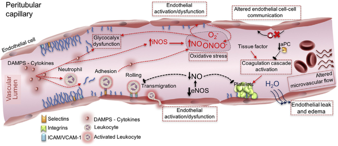
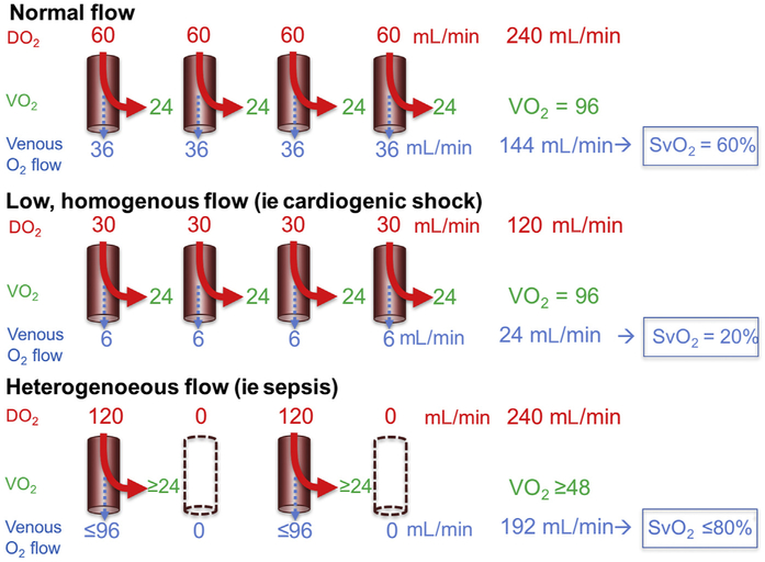
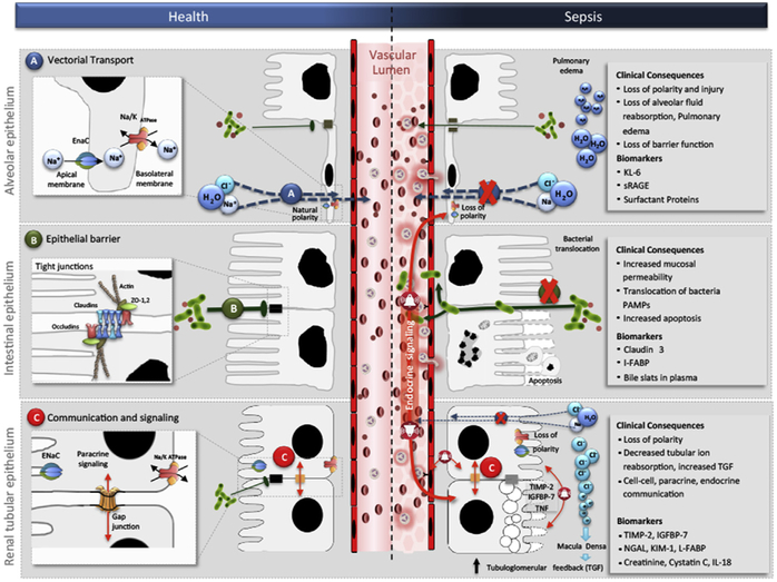
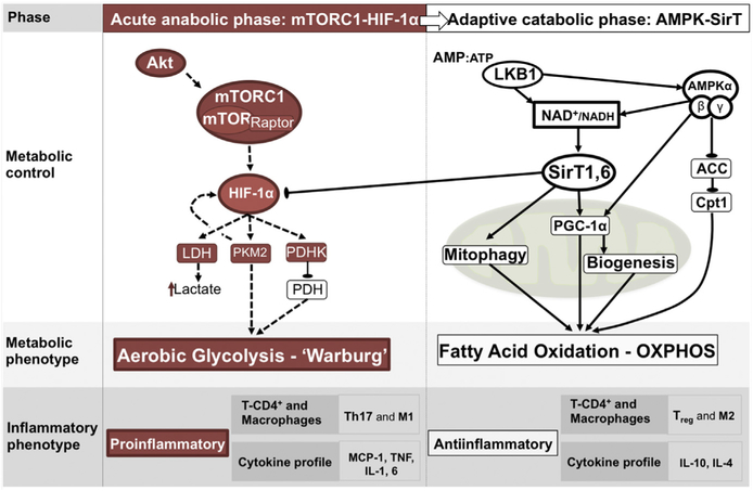
Sepsis-associated organ dysfunction involves multiple responses to inflammation, including endothelial and microvascular dysfunction, immune and autonomic dysregulation, and cellular metabolic reprogramming. The effect of targeting these mechanistic pathways on short- and long-term outcomes depends highly on the timing of therapeutic intervention. Furthermore, there is a need to understand the adaptive or maladaptive character of these mechanisms, to discover phase-specific biomarkers to guide therapy, and to conceptualize these mechanisms in terms of resistance and tolerance.

Keywords: Inflammation; Metabolism; Microcirculation; Mitochondria; Organ dysfunction; Sepsis.


Figures

Fig. 1.
Fig. 1.
Mechanisms leading to microcirculatory dysfunction: damage- and pathogen-associated molecular patterns (DAMPs and PAMPs), oxidative stress, and altered nitric oxide production contribute to endothelial dysfunction. As a result, glycocalyx denudation alters the colloid osmotic gradient between the capillary lumen and protein-rich area protected by the glycocalyx layer, leading to increased capillary leak and to increased adhesion of platelets and neutrophils. An inducible nitric oxide synthase (iNOS)–dependent decrease in endothelial nitric oxide synthase (eNOS)–derived nitric oxide production results in loss of endothelial protection via loss of direct vasodilation and loss of platelet aggregation and leukocyte activation inhibition and platelet adhesion and coagulation cascade activation in the setting of endothelial dysfunction. VCAM-1, vascular cell adhesion protein 1. (Adapted from Gómez H, Kellum JA. Sepsis-induced acute kidney injury. Curr Opin Crit Care 2016;22(6):546–53.)
Fig. 2.
Fig. 2.
Decreased efficiency of oxygen delivery with heterogeneous flow. In normal microcirculatory flow, cells extract oxygen to meet oxygen consumption (Vo2) requirements. Homogenously decreased flow results in decreased oxygen delivery (DO2) but preserved extraction and Vo2, with a resulting decrease in venous oxygen saturation (SvO2). In the presence of heterogeneous flow, despite preservation of total oxygen delivery, only a portion of capillaries is perfused. Cells too distant from perfused vessels do not receive enough oxygen to meet metabolic requirements and become hypoxic. Therefore, provided no changes in Vo2, hypoxic regions could be found in the presence of an elevated SvO2. (Modified from De Backer D, Ospina-Tascon G, Salgado D, et al. Monitoring the microcirculation in the critically ill patient: current methods and future approaches. Intensive Care Med 2010;36:1813–25; with permission.)
Fig. 3.
Fig. 3.
Common epithelial functions in health and in sepsis. Vectorial transport in the lung becomes altered in sepsis causing loss of polarity and barrier function (A); epithelial barrier function in the intestine becomes impaired in sepsis causing increased permeability and bacterial translocation (B); and communication and signaling in the kidney becomes associated with cell-cell, paracrine, and endocrine communication in sepsis (C). Cl, chloride; H2O, water; I-FABP, intestinal-fatty acid binding protein; IGFBP-7, insulinlike growth factor binding protein-7; IL-18, interleukin-18; K, potassium; KIM-1, kidney injury molecule-1; KL-6, Krebs Von den Lungen-6; Na, sodium; NGAL, neutrophil gelatinase–associated lipocalin; PAMPs, pathogen-associated molecular patterns; sRAGE, soluble receptor of advanced glycation end products; TGF, transforming growth factor; TIMP-2, tissue inhibitor of metalloproteinases-2; ZO, zonula occludens. (From Acute dialysis quality initiative 14. Available at: www.adqi.org. Accessed June 7, 2017; with permission.)
Fig. 4.
Fig. 4.
Inflammation-induced metabolic shift from OXPHOS to aerobic glycolysis. In monocytes, the shift toward aerobic glycolysis has been attributed to the activation of HIF-1 α. This shift results in increased expression of cytoplasm glucose transporters, enhanced activity of glycolytic enzymes, expression of pyruvate kinase isoform M2 (PKM2, slows conversion of phos-phoenolpyruvate to pyruvate), and expression of pyruvate dehydrogenase kinase (PDHK, limits pyruvate entrance into Krebs cycle). A shift back to OXPHOS has been attributed to the nicotinamide adenine dinucleotide (NAD)–dependent deacetylases (sirtuins) SirT1 and SirT6, which blocks the HIF-1α axis. ACC, acetyl CoA carboxylase; Akt, serine/threonine-specific protein kinase; AMP, adenosine monophosphate; ATP, adenosine triphosphate; cpt1, carnitine palmitoyl transferase; HIF-1a, hypoxia inducible factor-1 alpha; IL, interleukin; IL-1, IL-4, IL-6, and IL-10, interleukin 1, 4, 6 and 10, respectively; LDH, lactate dehydrogenase; M1, macrophage activation phenotype with inflammatory functions; M2, macrophage activation phenotype with anti-inflammatory functions; MCP-1, monocyte chemoattractant protein-1; mTOR, mammalian target of rapamicin; mTORC1, mammalian target of rapamicin complex; NAD+/NADH, oxidized/reduced nicotinamide adenine dinucleotide; PDH, pyruvate dehydrogenase; PGC-1a, Peroxisome proliferator-activated receptor gamma coactivator 1-alpha; Sirt1,6, sirtuin 1 and 6; T-CD4+, T lymphocyte-cluster of differentiation 4; Th17, T helper 17 cell; TNF, tumor necrosis factor; Treg, regulatory T cell. (Adapted from Gómez H, Kellum JA, Ronco C, et al. Metabolic reprogramming and tolerance during sepsis-induced AKI. Nat Rev Nephrol 2017;13(3):143–51.)
Fig. 5.
Fig. 5.
Inflammation-induced metabolic reprogramming. Exposure of tubular epithelial cells to damage-associated molecular patterns (DAMPs) and pathogen-associated molecular patterns (PAMPs) leads to changes in metabolic regulation within the cell that can impact cell survival, organ function and, possibly, repair events after injury subsides. The image shows 3 possible domains of acute-phase metabolic regulation, including triggering of mitochondrial quality control processes including mitophagy and biogenesis, shifting metabolism from OXPHOS toward glycolysis, and inducing cell cycle arrest. FAO, fatty acid oxidation; IGFBP7, insulinlike growth factor-binding protein 7; OXPHOS, oxidative phosphorylation; TIMP-2, tissue inhibitor of metalloproteinases-2; TLR-4, toll-like receptor-4. (Adapted from Gómez H, Kellum JA. Sepsis-induced acute kidney injury. Curr Opin Crit Care 2016;22(6):546–53.)

References

    1. Singer M, Deutschman CS, Seymour CW, et al. The third international consensus definitions for sepsis and septic shock (sepsis-3). JAMA 2016;315: 801–10. - PMC - PubMed
    1. Angus DC, van der Poll T. Severe sepsis and septic shock. N Engl J Med 2013; 369:840–51. - PubMed
    1. Yende S, Iwashyna TJ, Angus DC. Interplay between sepsis and chronic health. Trends Mol Med 2014;20:234–8. - PMC - PubMed
    1. Langenberg C, Wan L, Egi M, et al. Renal blood flow in experimental septic acute renal failure. Kidney Int 2006;69:1996–2002. - PubMed
    1. Prowle JR, Ishikawa K, May CN, et al. Renal blood flow during acute renal failure in man. Blood Purif 2009;28:216–25. - PubMed

MeSH terms