. 2017 Dec 29;292(52):21282-21290.
doi: 10.1074/jbc.C117.815845. Epub 2017 Nov 17.

Functional analyses of a human vascular tumor FOS variant identify a novel degradation mechanism and a link to tumorigenesis

Affiliations

Functional analyses of a human vascular tumor FOS variant identify a novel degradation mechanism and a link to tumorigenesis

David G P van IJzendoorn et al. J Biol Chem. .

Abstract

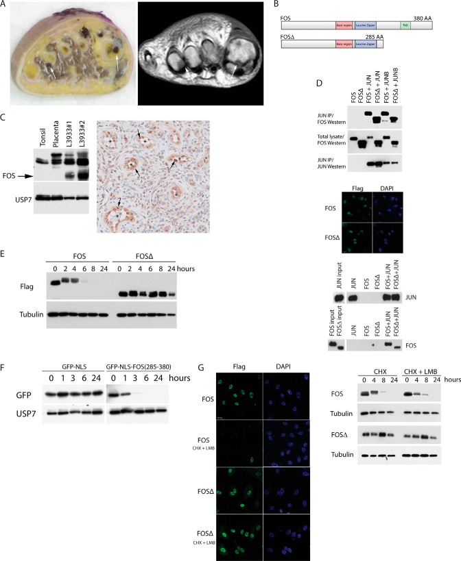
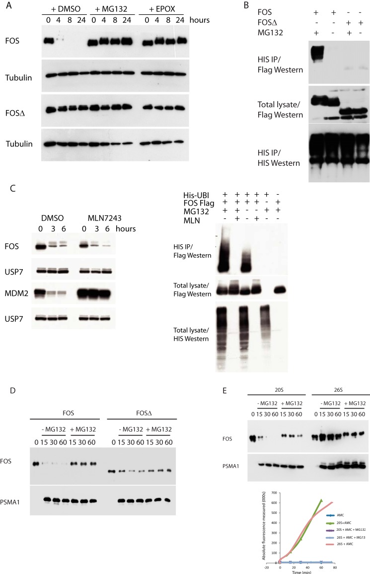
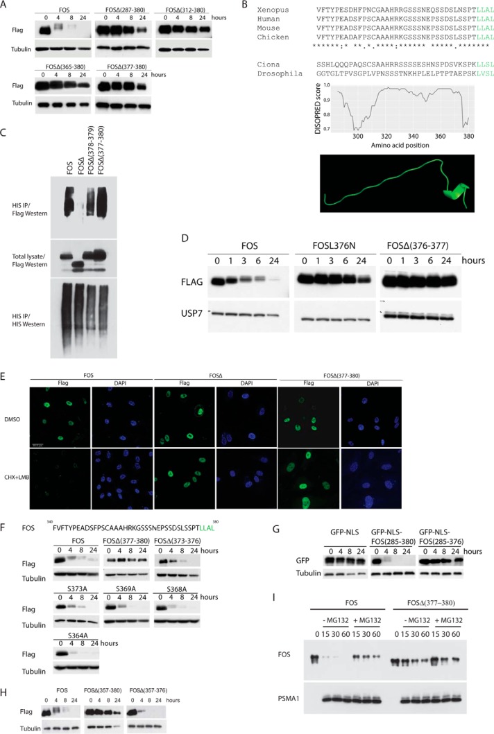
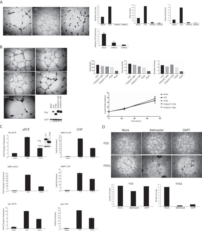
Epithelioid hemangioma is a locally aggressive vascular neoplasm, found in bones and soft tissue, whose cause is currently unknown, but may involve oncogene activation. FOS is one of the earliest viral oncogenes to be characterized, and normal cellular FOS forms part of the activator protein 1 (AP-1) transcription factor complex, which plays a pivotal role in cell growth, differentiation, and survival as well as the DNA damage response. Despite this, a causal link between aberrant FOS function and naturally occurring tumors has not yet been established. Here, we describe a thorough molecular and biochemical analysis of a mutant FOS protein we identified in these vascular tumors. The mutant protein lacks a highly conserved helix consisting of the C-terminal four amino acids of FOS, which we show is indispensable for fast, ubiquitin-independent FOS degradation via the 20S proteasome. Our work reveals that FOS stimulates endothelial sprouting and that perturbation of normal FOS degradation could account for the abnormal vessel growth typical of epithelioid hemangioma. To the best of our knowledge, this is the first functional characterization of mutant FOS proteins found in tumors.

Keywords: angiogenesis; c-Fos; cancer biology; protein degradation; transcription regulation; vascular biology.


Conflict of interest statement

The authors declare that they have no conflicts of interest with the contents of this article

Figures

Figure 1.
Figure 1.
A, epithelioid hemangioma case L3933. Left panel, gross specimen with polyostotic localization of a hemorrhagic tumor in the 1st and 4th metatarsal bones of the foot (arrows). Right panel, corresponding T1 weighted MR image. B, tumor FOSΔ lacks the C-terminal 95 amino acids (including the C-terminal TAD). IP, immunoprecipitation. C, left panel, Western blot of endogenous FOS proteins in control tonsil and placenta cell lysates compared with epithelioid hemangioma tumor cell lysates. Mutant FOSΔ protein is highlighted with an arrow. Right panel, high FOS expression (arrows) is indicated in the endothelial cells of epithelioid hemangioma tumor blood vessels (*). D, AP-1 heterodimers were immunopurified from cells transfected with the indicated constructs (top panel). Immunofluorescence shows both FOS and FOSΔ localize to the nucleus (middle panel). FOS (and FOSΔ), JUN heterodimers bind to consensus AP-1 DNA-binding sites (bottom panel). E, FOS stability assay on HUVECs stably expressing FOS or FOSΔ. F, protein stability assay on HUVECs stably expressing either GFP or a GFP-FOS fusion (encompassing the C-terminal 95 amino acids of FOS). G, HUVECs expressing the indicated proteins were incubated with or without leptomycin B (LMB) in the presence of cycloheximide (CHX). Left panel, immunofluorescence. Right panel, Western blots.
Figure 2.
Figure 2.
A, FOS stability assay on HUVECs stably expressing FOS or FOSΔ. B, ubiquitin assay of cells transfected with the indicated constructs together with 10× HIS epitope-tagged ubiquitin. C, left panel, FOS stability assay on HUVECs stably expressing FOS in the presence or absence of MLN7243. Right panel, ubiquitin assay of cells transfected with the indicated constructs and cultured in the presence or absence of MG132 and MLN7243. IP, immunoprecipitation. D, in vitro translated FOS proteins were incubated with purified 20S proteasomes for the shown time course (minutes). 20S protein levels were determined by Western blotting using an antibody directed against PSMA1. 20S proteasome activity was independently quantified using the suc-Leu-Leu-Val-Tyr-AMC peptide (as shown in E). E, experiment performed as in D.
Figure 3.
Figure 3.
A, FOS stability assay on HUVECs stably expressing the indicated FOS deletion mutants. B, ab initio modeling of the FOS C terminus. C, ubiquitin assay performed on cells transfected with the indicated constructs together with 10× HIS epitope-tagged ubiquitin. Cells were cultured in the presence of MG132. IP, immunoprecipitation. D, FOS stability assay on HUVECs stably expressing the indicated FOS deletion mutants. E, HUVECs expressing the indicated proteins were incubated with or without leptomycin B (LMB) in the presence of cycloheximide (CHX). FOS was visualized by immunofluorescence. F, FOS stability assay on HUVECs stably expressing the indicated FOS deletion mutants. G, protein stability assay on HUVECs stably expressing either GFP, a GFP-FOS fusion (encompassing the C-terminal 95 amino acids of FOS), or the same fusion lacking the last four amino acids of FOS. H, FOS stability assay on HUVECs stably expressing the indicated FOS deletion mutants. FOSΔ(357–380) lacks the C-terminal 23 amino acids (the IDR). FOSΔ(357–376) lacks the IDR but retains the C-terminal four amino acids. I, in vitro FOS stability assay as described in Fig. 2, D and E.
Figure 4.
Figure 4.
A, HUVECs lacking endogenous FOS or ectopically expressing wild-type FOS were grown on Matrigel. A representative of several independent experiments is shown. Sprouting was quantified after 24 h using in-house computer software. Loss of FOS was determined by qPCR (lowermost graph). B, Matrigel sprouting assay (see A) on HUVECs stably expressing the indicated FOS proteins. Lower graph, cell proliferation assay of the same HUVECs lines. Triplicate measurements were made at each time point. Values are means ± S.E. of the mean. C, left panel, expression levels of the indicated transcripts in HUVECs were determined by real-time qPCR. All values were averaged relative to TATA-binding protein (TBP), signal recognition particle receptor (SRPR), and calcium-activated neutral proteinase 1 (CAPNS1). Values were normalized against mock-treated cells. Values represent ± S.D. (n = 3). Right panel, a ChIP analysis of FOS association with the indicated promoters in HUVECs stably expressing FOS or tumor FOSΔ. Three different primer sets were used for each promoter region. A single representative is shown (all three gave similar results). Results are presented as mean fold changes in recovery (as a fraction of input) relative to the Mock infected cells. Error bars represent the standard deviation (n = 3). Relative FOS and FOSΔ protein levels were determined by Western blotting. D, HUVECs stably expressing the indicated FOS proteins were grown on Matrigel in the presence or absence of the MMP inhibitor, batimastat (10 μm), or the γ-secretase inhibitor, DAPT (10 μm). Sprouting was quantified after 48 h.

References

    1. Rosenberg A. E., and Bovée J. V. (2013) in WHO Classification of Tumours of Soft Tissue and Bone (Fletcher C. D., Bridge J. A., Hogendoorn P. C., and Mertens F., eds) pp. 333–334, IARC Press, Lyon, France
    1. van IJzendoorn D. G., de Jong D., Romagosa C., Picci P., Benassi M. S., Gambarotti M., Daugaard S., van de Sande M., Szuhai K., and Bovée J. V. (2015) Fusion events lead to truncation of FOS in epithelioid hemangioma of bone. Genes Chromosomes Cancer 54, 565–574 - PubMed
    1. Huang S. C., Zhang L., Sung Y. S., Chen C. L., Krausz T., Dickson B. C., Kao Y. C., Agaram N. P., Fletcher C. D., and Antonescu C. R. (2015) Frequent FOS gene rearrangements in epithelioid hemangioma: A molecular study of 58 cases with morphological reappraisal. Am. J. Surg. Pathol. 39, 1313–1321 - PMC - PubMed
    1. Wagner E. F. (2001) AP-1 reviews. Oncogene 20, 2333–2497
    1. Johnson R. S., Spiegelman B. M., and Papaioannou V. (1992) Pleiotropic effects of a null mutation in the c-fos proto-oncogene. Cell 71, 577–586 - PubMed

Publication types

MeSH terms

Substances