. 2018 Jan 19;293(3):1007-1017.
doi: 10.1074/jbc.M117.805960. Epub 2017 Nov 17.

The prototypical proton-coupled oligopeptide transporter YdgR from Escherichia coli facilitates chloramphenicol uptake into bacterial cells

Affiliations

The prototypical proton-coupled oligopeptide transporter YdgR from Escherichia coli facilitates chloramphenicol uptake into bacterial cells

Bala K Prabhala et al. J Biol Chem. .

Abstract

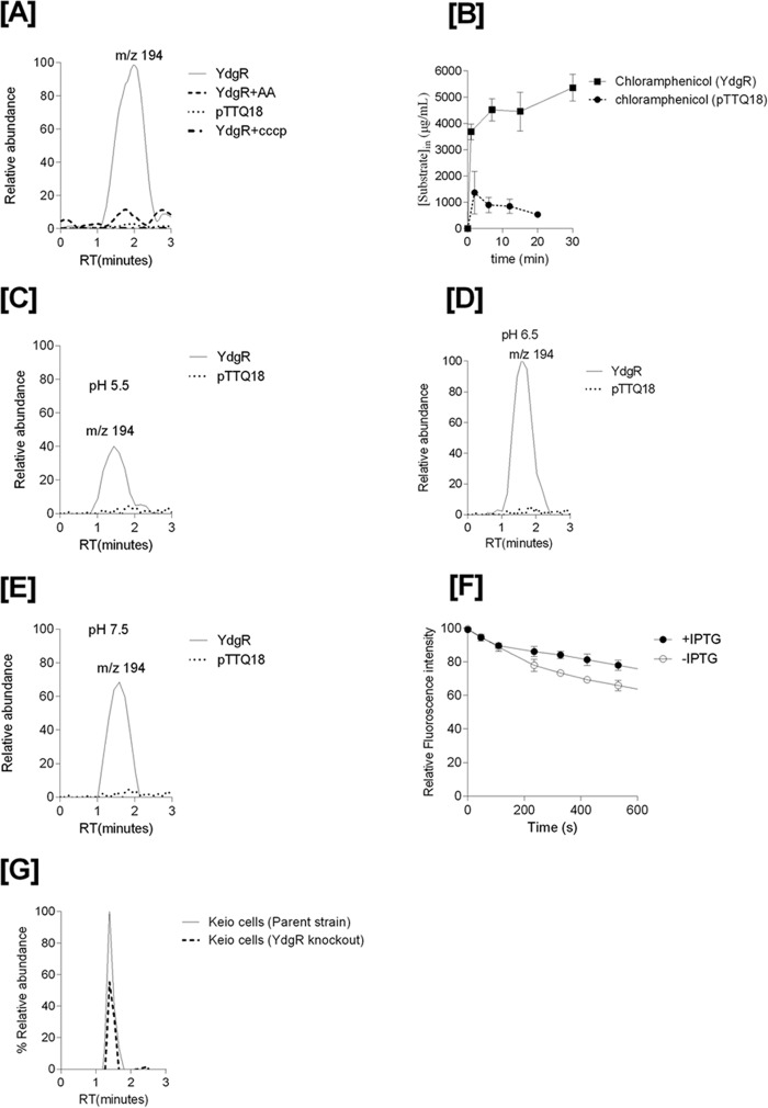
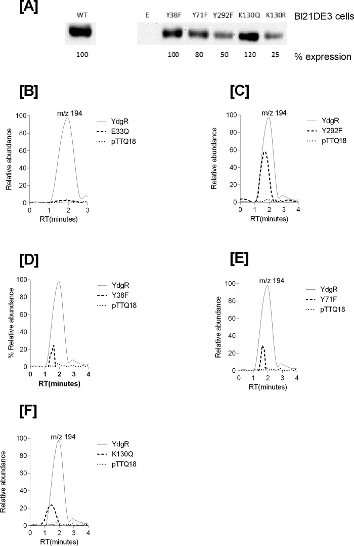
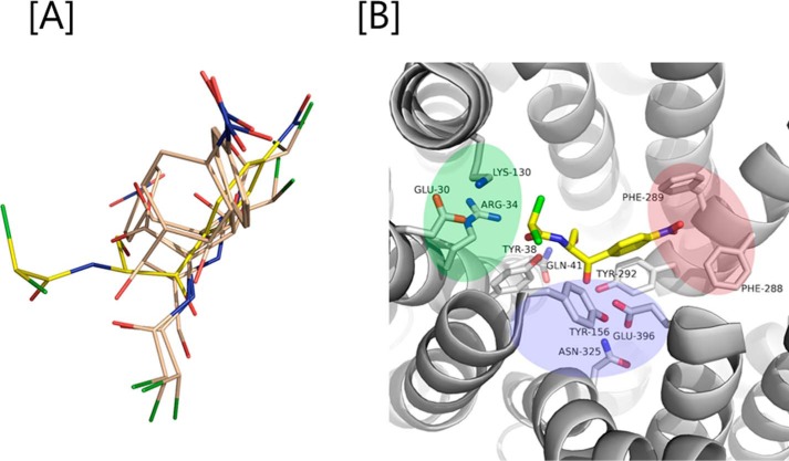
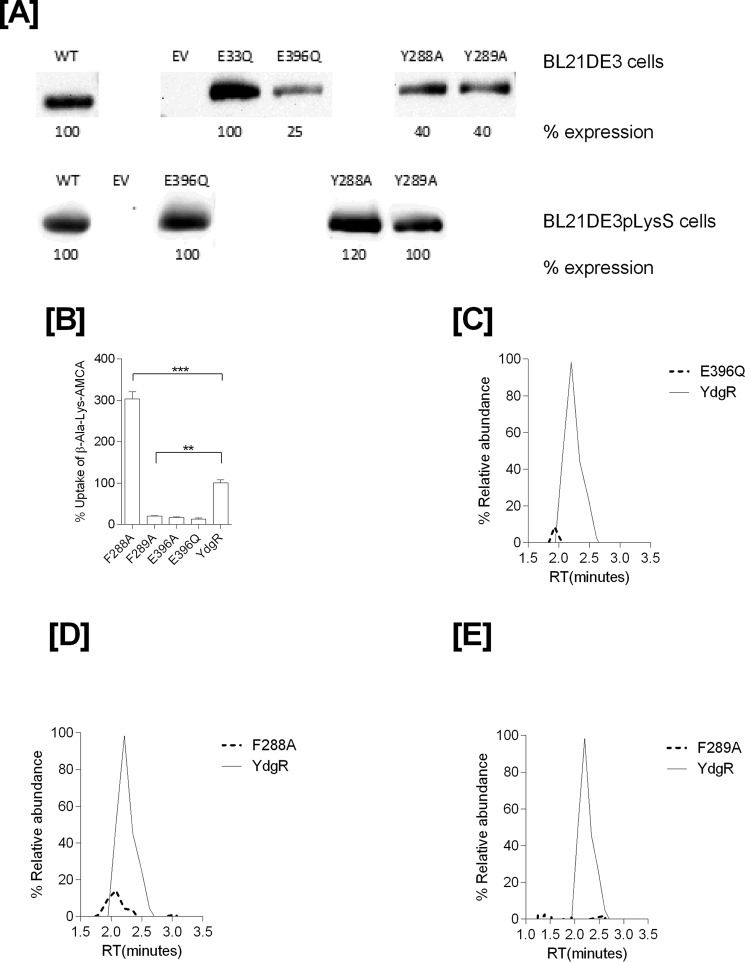
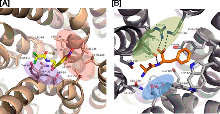
Chloramphenicol (Cam) is a broad-spectrum antibiotic used to combat bacterial infections in humans and animals. Cam export from bacterial cells is one of the mechanisms by which pathogens resist Cam's antibacterial effects, and several different proteins are known to facilitate this process. However, to date no report exists on any specific transport protein that facilitates Cam uptake. The proton-coupled oligopeptide transporter (POT) YdgR from Escherichia coli is a prototypical member of the POT family, functioning in proton-coupled uptake of di- and tripeptides. By following bacterial growth and conducting LC-MS-based assays we show here that YdgR facilitates Cam uptake. Some YdgR variants displaying reduced peptide uptake also exhibited reduced Cam uptake, indicating that peptides and Cam bind YdgR at similar regions. Homology modeling of YdgR, Cam docking, and mutational studies suggested a binding mode that resembles that of Cam binding to the multidrug resistance transporter MdfA. To our knowledge, this is the first report of Cam uptake into bacterial cells mediated by a specific transporter protein. Our findings suggest a specific bacterial transporter for drug uptake that might be targeted to promote greater antibiotic influx to increase cytoplasmic antibiotic concentration for enhanced cytotoxicity.

Keywords: antibiotics; membrane transport; molecular docking; site-directed mutagenesis; transporter.


Conflict of interest statement

The authors declare that they have no conflicts of interest with the contents of this article

Figures

Figure 1.
Figure 1.
A, LC-MS electrospray ion-trap spectrum of chloramphenicol standard (15 μg/ml) dissolved in cell lysates. The parent ion at m/z 321 is observed but of low intensity. The most prominent product ion at m/z 194 was monitored in the uptake assays. B, β-Ala-Lys(AMCA) (0.5 mm) uptake and inhibition in the presence of Cam (0.5 mm) as a competitor (p value < 0.05, n = 3).
Figure 2.
Figure 2.
Growth curves of YdgR, pTTQ18 vector, and YdgR–E33Q mutant in BL21(DE3)pLysS cells (in the presence of 100 μg/ml of ampicillin and 34 μg/ml of chloramphenicol). Arrow marks indicates time of induction with IPTG. Closed symbols for the cells induced with IPTG and open symbols for cells without IPTG. A, YdgR; B, pTTQ18 vector; and C, YdgR–E33Q (the insert of Western blot bands are spliced from the same gel). Open squares indicate the growth of BL21(DE3) cells in the presence of 100 μg/ml of ampicillin and absence of 34 μg/ml of chloramphenicol. D, YdgR; n = 3.
Figure 3.
Figure 3.
MIC determination of YdgR and pTTQ18 (pTTQ18 vector) expressing BL21(DE3)pLysS cells and BL21DE3. A, plates supplemented with 0.1 mm IPTG. B, plates without IPTG induction. n = 3.
Figure 4.
Figure 4.
A, mass chromatograms of chloramphenicol detected in the BL21(DE3) cell lysates of YdgR (thin line), pTTQ18 vector (small dots), YdgR in competition with Ala–Ala (broken lines). B, time-dependent uptake of chloramphenicol. The optimal uptake of YdgR is at pH 6.5 (100%) and C and E are shown relative to D. C, uptake of chloramphenicol at pH 5.5. D, uptake of chloramphenicol at pH 6.5. E, uptake of chloramphenicol at pH 7.5, n = 3. F, ethidium bromide efflux in the presence (closed circles) and absence (pTTQ18, circles) of YdgR. G, mass chromatogram of chloramphenicol in an E. coli parent strain (thin line) and YdgR deletion strain (broken lines). n = 3.
Figure 5.
Figure 5.
A, Western blots showing expression of YdgR–mutants in BL21(DE3) cells. B, mass chromatograms showing expression normalized uptake of chloramphenicol in the BL21(DE3) cell in expressing YdgR (thin line), pTTQ18 vector (small dots), YdgR–E33Q (broken lines). C, YdgR (thin line), pTTQ18 vector (small dots), YdgR–Y292F (broken lines). D, YdgR (thin line), pTTQ18 vector (small dots), YdgR–Y38F (broken lines). E, YdgR (thin line), pTTQ18 vector (small dots), YdgR–Y71F (broken lines). F, YdgR (thin line), pTTQ18 vector (small dots), YdgR–K130Q (broken lines). n = 3.
Figure 6.
Figure 6.
A, various conformations of Cam (gray color; one in yellow is the most preferred confirmation) out by docking it into the binding pocket YdgR. B, green, blue, and magenta colors indicate protonation, hydrophilic, and hydrophobic regions in the active site. Interactions between Cam (yellow color) and YdgR (white color) from the docking pose.
Figure 7.
Figure 7.
Expression and functional characterization of mutants prepared based on predicted docking of Cam at the active site. A, Western blots of mutants expressed in BL21(DE3) and BL21(DE3)pLysS cells. B, β-Ala-Lys(AMCA) (0.5 mm) uptake profiles of ydgR/ydgR–mutants. Mass chromatograms showing expression normalized uptake of chloramphenicol in the BL21(DE3) cell lysates. Broken lines represent mutants and thin solid line represent wild type YdgR. C, YdgR (thin line), YdgR–E396Q (broken lines). D, YdgR (thin line), YdgR–F288A (broken lines). E, YdgR (thin line), YdgR–F289A (broken lines). n = 3.
Figure 8.
Figure 8.
The red, blue, and green color patches represent the hydrophobic, hydrophilic, and protonation sites, respectively. A, interactions between Cam (yellow color) and MdfA (wheat color) in a crystal structure (PDB code 4ZOW). B, interactions between the Ala–Phe peptide (yellow color) and PepTSt (gray color) in the crystal structure (PDB code 5D59).

References

    1. Sutcliffe J. A. (2005) Improving on nature: antibiotics that target the ribosome. Curr. Opin. Microbiol. 8, 534–542 10.1016/j.mib.2005.08.004 - DOI - PubMed
    1. Franje C. A., Chang S. K., Shyu C. L., Davis J. L., Lee Y. W., Lee R. J., Chang C. C., and Chou C. C. (2010) Differential heat stability of amphenicols characterized by structural degradation, mass spectrometry and antimicrobial activity. J. Pharm. Biomed. Anal. 53, 869–877 - PubMed
    1. Gruhzit O. M., et al. , (1949) Chloramphenicol (chloromycetin), an antibiotic. pharmacological and pathological studies in animals. J. Clin. Invest. 28, 943–952 - PMC - PubMed
    1. Hanekamp J. C., and Bast A. (2015) Antibiotics exposure and health risks: chloramphenicol. Environ. Toxicol. Pharmacol. 39, 213–220 10.1016/j.etap.2014.11.016 - DOI - PubMed
    1. Mortimer P. G., and Piddock L. J. (1993) The accumulation of five antibacterial agents in porin-deficient mutants of Escherichia coli. J. Antimicrob. Chemother. 32, 195–213 10.1093/jac/32.suppl_B.195, 10.1093/jac/32.2.195 - DOI - DOI - PubMed

Publication types

MeSH terms

LinkOut - more resources