Review
. 2017 Dec 15;45(6):1313-1321.
doi: 10.1042/BST20170095. Epub 2017 Nov 17.

Distinct RNA-unwinding mechanisms of DEAD-box and DEAH-box RNA helicase proteins in remodeling structured RNAs and RNPs

Affiliations
Review

Distinct RNA-unwinding mechanisms of DEAD-box and DEAH-box RNA helicase proteins in remodeling structured RNAs and RNPs

Benjamin Gilman et al. Biochem Soc Trans. .

Abstract

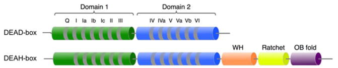
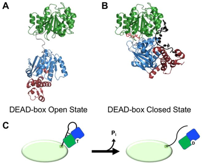
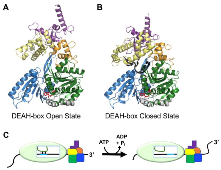
Structured RNAs and RNA-protein complexes (RNPs) fold through complex pathways that are replete with misfolded traps, and many RNAs and RNPs undergo extensive conformational changes during their functional cycles. These folding steps and conformational transitions are frequently promoted by RNA chaperone proteins, notably by superfamily 2 (SF2) RNA helicase proteins. The two largest families of SF2 helicases, DEAD-box and DEAH-box proteins, share evolutionarily conserved helicase cores, but unwind RNA helices through distinct mechanisms. Recent studies have advanced our understanding of how their distinct mechanisms enable DEAD-box proteins to disrupt RNA base pairs on the surfaces of structured RNAs and RNPs, while some DEAH-box proteins are adept at disrupting base pairs in the interior of RNPs. Proteins from these families use these mechanisms to chaperone folding and promote rearrangements of structured RNAs and RNPs, including the spliceosome, and may use related mechanisms to maintain cellular messenger RNAs in unfolded or partially unfolded conformations.

Keywords: RNA structure; helicase; self-splicing intron; splicing; unwinding.


Conflict of interest statement

Competing Interests

The authors declare that there are no competing interests associated with the manuscript.

Figures

Figure 1
Figure 1
Structural arrangement of conserved domains in DEAD-box and DEAH-box helicases. The protein families share two conserved, RecA-like domains with numbered sequence motifs that are highly conserved within each family. Many DEAD-box proteins have additional N- and C-terminal extensions that are required for their specific functions but are not conserved between different DEAD-box proteins and are not shown here. In contrast, DEAH-box proteins share conserved C-terminal domains consisting of winged helix (WH), ratchet-like, and oligosaccharide binding fold (OB fold) domains.
Figure 2
Figure 2
Remodeling of exposed RNA duplexes by DEAD-box proteins. (A) Structural model for the open conformation of a DEAD-box protein (S. cerevisiae Mss116) prior to substrate binding (16). Conserved domains 1 and 2 are colored as in Figure 1. Mss116 and its homologs have an additional C-terminal extension (magenta) that is not present in all DEAD-box proteins. (B) Crystal structure representing the closed state of a DEAD-box protein (Mss116) following strand displacement (PDB: 3I5X) (20). The bound ssRNA strand is shown in black and the bound AMP-PNP molecule is red. (C) Model for unwinding of an exposed RNA duplex from a larger RNA or RNP by a DEAD-box helicase (shown as green and blue D1 and D2 respectively). The ATP-bound DEAD-box protein interacts with an exposed RNA helix on the surface of a structured RNA or RNP (green oval). Protein binding results in unwinding of the helix, allowing the partner strands to form new inter- or intramolecular interactions. ATP hydrolysis and release of Pi then allow the DEAD-box protein to release from the liberated ssRNA.
Figure 3
Figure 3
Remodeling of internal RNA duplexes by DEAH-box proteins. (A) Crystal structure of Prp43 with bound ADP-BeFx showing the open conformation, which facilitates RNA loading (PDB: 5LTK)(57). Domains are colored as in Fig. 1. (B) Crystal structure of the closed conformation of Prp43. The structure includes bound ADP-BeFx and U7 RNA and reflects the conformation that follows RNA loading (PDB: 5LTA)(57). (C) Model for DEAH-box helicase-catalyzed disruption of an RNA duplex within a larger RNP complex by molecular winching (68). Translocation of the protein against the RNP surface results in movement of the RNA (black) relative to the interior of the RNAP. Because its base-pairing partner (green) is held in place relative to the RNP, the movement of the black RNA results in disruption of the base pairs between the two RNAs.

References

    1. Herschlag D. RNA chaperones and the RNA folding problem. J Biol Chem. 1995;270(36):20871–4. - PubMed
    1. Karpel RL, Swistel DG, Miller NS, Geroch ME, Lu C, Fresco JR. Acceleration of RNA renaturation by nucleic acid unwinding proteins. Brookhaven Symp Biol. 1975;(26):165–74. - PubMed
    1. Russell R. RNA misfolding and the action of chaperones. Front Biosci. 2008;13:1–20. - PMC - PubMed
    1. Millevoi S, Moine H, Vagner S. G-quadruplexes in RNA biology. Wiley Interdiscip Rev RNA. 2012;3(4):495–507. - PubMed
    1. Wan Y, Kertesz M, Spitale RC, Segal E, Chang HY. Understanding the transcriptome through RNA structure. Nat Rev Genet. 2011;12(9):641–55. - PMC - PubMed

LinkOut - more resources