. 2017 Dec 5;114(49):12994-12999.
doi: 10.1073/pnas.1705165114. Epub 2017 Nov 17.

Cancer cells induce interleukin-22 production from memory CD4+ T cells via interleukin-1 to promote tumor growth

Affiliations

Cancer cells induce interleukin-22 production from memory CD4+ T cells via interleukin-1 to promote tumor growth

Cornelia Voigt et al. Proc Natl Acad Sci U S A. .

Abstract

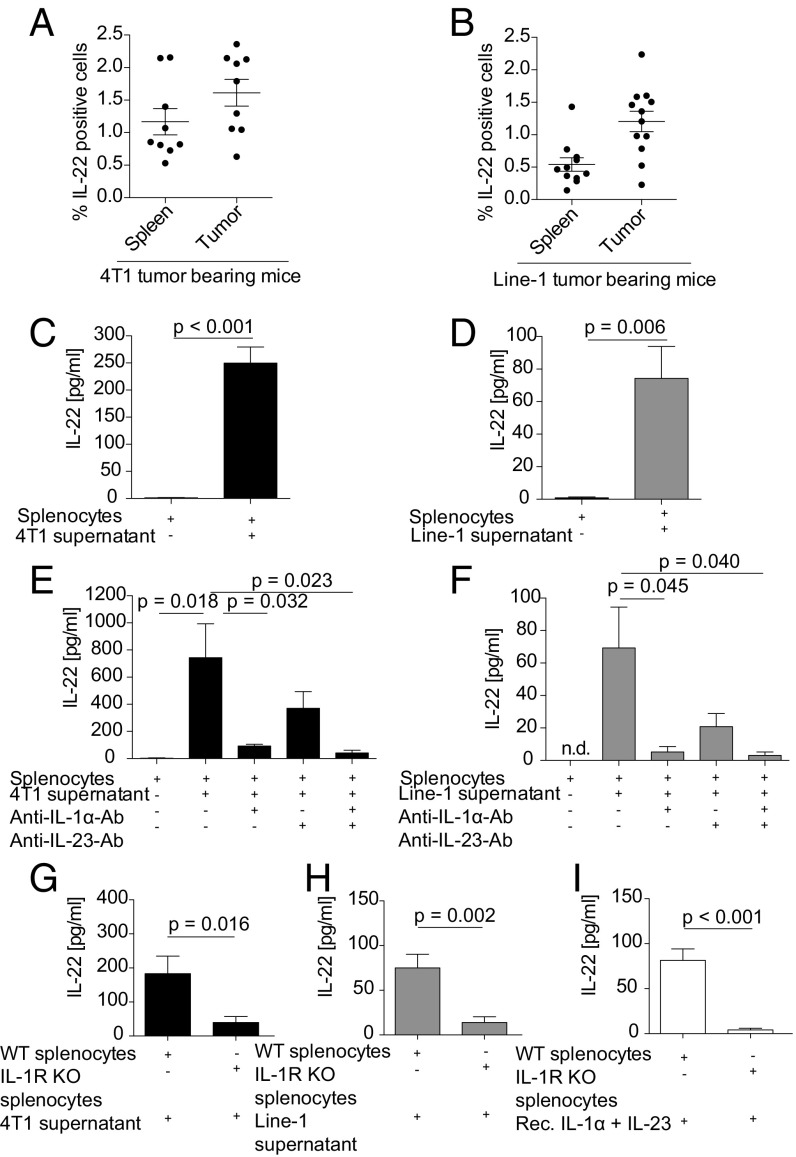
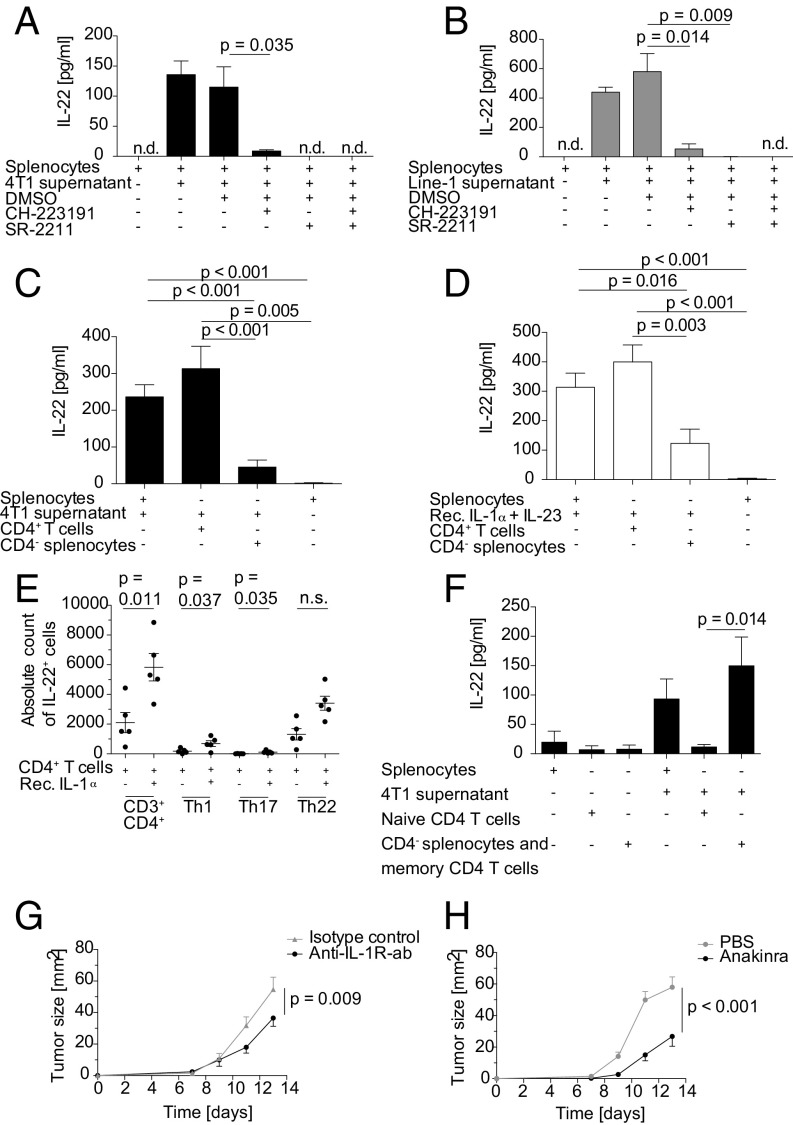
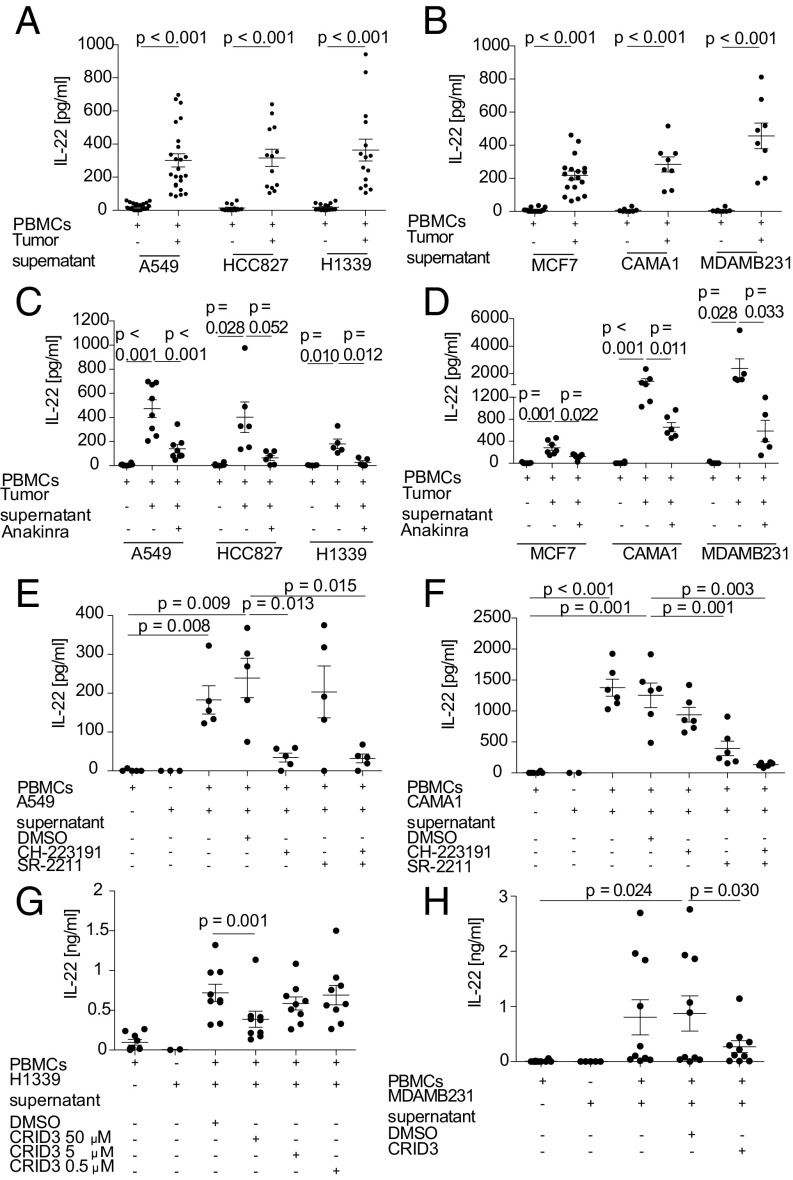
IL-22 has been identified as a cancer-promoting cytokine that is secreted by infiltrating immune cells in several cancer models. We hypothesized that IL-22 regulation would occur at the interface between cancer cells and immune cells. Breast and lung cancer cells of murine and human origin induced IL-22 production from memory CD4+ T cells. In the present study, we found that IL-22 production in humans is dependent on activation of the NLRP3 inflammasome with the subsequent release of IL-1β from both myeloid and T cells. IL-1 receptor signaling via the transcription factors AhR and RORγt in T cells was necessary and sufficient for IL-22 production. In these settings, IL-1 induced IL-22 production from a mixed T helper cell population comprised of Th1, Th17, and Th22 cells, which was abrogated by the addition of anakinra. We confirmed these findings in vitro and in vivo in two murine tumor models, in primary human breast and lung cancer cells, and in deposited expression data. Relevant to ongoing clinical trials in breast cancer, we demonstrate here that the IL-1 receptor antagonist anakinra abrogates IL-22 production and reduces tumor growth in a murine breast cancer model. Thus, we describe here a previously unrecognized mechanism by which cancer cells induce IL-22 production from memory CD4+ T cells via activation of the NLRP3 inflammasome and the release of IL-1β to promote tumor growth. These findings may provide the basis for therapeutic interventions that affect IL-22 production by targeting IL-1 activity.

Keywords: anakinra; cancer immunology; inflammasome; interleukin-1; interleukin-22.


Conflict of interest statement

The authors declare no conflict of interest.

Figures

Fig. 1.
Fig. 1.
Murine lung and breast cancer cell lines induce IL-22 from splenocytes via tumor-derived IL-1α. (A and B) Single-cell suspensions of 4T1 (A) and Line-1 (B) s.c. tumors were analyzed by flow cytometry for total intracellular IL-22 expression. Values in A and B represent pooled data of three independent experiments with three mice per group and four independent experiments with three mice per group, respectively. (C and D) Splenocytes (2 × 10⁶/mL) were stimulated with 50% cell-free 4T1 (C) or Line-1 (D) tumor-cell supernatant for 6 d. Mean values from five independent experiments are shown. (E and F) Splenocytes were stimulated with 4T1 (E) or Line-1 (F) tumor-cell supernatant in the presence or absence of anti–IL-1α or anti–IL-23 blocking antibodies (2.5 µg/mL) or both for 6 d. The mean values of a minimum of four independent experiments are shown. (GI) Wild-type or IL-1R knock-out splenocytes were stimulated with 4T1 supernatant (G), Line-1 supernatant (H), or 20 ng/mL recombinant IL-1α and IL-23, respectively (I). Values in GI represent pooled data of two independent experiments with four to seven mice per group. IL-22 production was quantified by ELISA (CI). Error bars represent the SEM; P values from a two-sided Student’s t test are shown; n.d., not detectable; rec., recombinant.
Fig. 2.
Fig. 2.
IL-22 is secreted from memory CD4+ T cells in an AhR- and RORγt-dependent manner, and in vivo neutralization of IL-1 reduces tumor growth. (A and B) Splenocytes (2 × 10⁶/mL) were stimulated with 4T1 supernatant (A) or Line-1 supernatant (B) in the presence or absence of 10 µM CH-223191 (AhR antagonist) or 5 µM SR-2211 (RORγt antagonist) for 6 d. Values in A are the mean of three different experiments performed in triplicate. Values in B are representative of five different experiments performed in triplicate. IL-22 production was quantified by ELISA. (C and D) MACS-enriched CD4+ T cells and CD4 splenocytes or total splenocytes were stimulated with 4T1 tumor supernatant (C) or 100 ng/mL recombinant IL-1α and IL-23 (D) for 6 d. Mean values of a minimum of four independent experiments are shown. (E) MACS-enriched CD4+ T cells were stimulated with 20 ng/mL recombinant IL-1α for 4 d. IL-22 production by Th1 (CD3+CD4+IFN-γ+), Th17 (CD3+CD4+IFN-γIL-17+), and Th22 (CD3+CD4+IFN-γIL-17) T cells was analyzed by flow cytometry (data represent two independent experiments with five different mice). (F) MACS-enriched naive CD4+ T cells and CD4 splenocytes including CD4+ CD44+ memory T cells were stimulated with 4T1 supernatant for 6 d. Mean values of three different experiments with eight replicates of supernatants are shown. (G) BALB/c mice bearing 1.25 × 10⁵ 4T1 tumor cells s.c. (n = 10 mice per group) were treated i.p. with 300 µg anti-mouse IL-1R antibody or isotype control every second day beginning on day 0. (H) C57BL/6 mice were injected s.c. in the right flank with 2.5 × 10⁵ E0771 tumor cells (n = 15 mice per group). Mice were treated with 1 mg anakinra or PBS i. p. every day beginning on day 0. In AF error bars represent the SEM, and P values by two-sided Student’s t test are shown. In G and H, statistical significance was analyzed by two-way ANOVA with correction for multiple testing; n.d., not detectable; n.s., not significant; rec., recombinant.
Fig. 3.
Fig. 3.
Human lung and breast tumor supernatants activate the NLRP3 inflammasome to release IL-1 and induce IL-22 in PBMCs from healthy donors in an AhR- and RORγT-dependent manner. (AD) PBMCs (2 × 10⁶/mL) were stimulated with 50% cell-free human lung cancer cell (A549, HCC827, H1339) conditioned supernatant (A) or breast cancer cell (MCF7, CAMA-1, MDAMB231) conditioned supernatant (B) for 6 d in the presence or absence of anakinra (500 ng/mL) (C and D). (E and F) PBMCs were stimulated with A549-conditioned (E) or CAMA-1–conditioned (F) tumor-cell supernatant for 6 d in the presence or absence of 10 µM CH-223191 or 5 µM SR-2211. (G and H) PBMCs were stimulated with H1339-conditioned (G) or MDAMB231-conditioned (H) tumor-cell supernatant for 6 d in the presence or absence of 50, 5, or 0.5 µM (H1339) or 50 µM (MDAMB231) CRID3. IL-22 production was quantified by ELISA (AH). n = 8–24 different donors in A and B, 5–8 different donors in C and D, 5–6 different donors in E and F, 9 different donors in G, and 10 different donors in H. Each dot in the graphs represents one donor. Error bars represent the SEM; P values from a two-sided Student’s t test are shown.

References

    1. Lim C, Savan R. The role of the IL-22/IL-22R1 axis in cancer. Cytokine Growth Factor Rev. 2014;25:257–271. - PubMed
    1. Hanahan D, Weinberg RA. Hallmarks of cancer: The next generation. Cell. 2011;144:646–674. - PubMed
    1. Sonnenberg GF, Fouser LA, Artis D. Border patrol: Regulation of immunity, inflammation and tissue homeostasis at barrier surfaces by IL-22. Nat Immunol. 2011;12:383–390. - PubMed
    1. Huber S, et al. IL-22BP is regulated by the inflammasome and modulates tumorigenesis in the intestine. Nature. 2012;491:259–263. - PMC - PubMed
    1. Kirchberger S, et al. Innate lymphoid cells sustain colon cancer through production of interleukin-22 in a mouse model. J Exp Med. 2013;210:917–931. - PMC - PubMed

Publication types

MeSH terms