. 2017 Nov 17;7(1):15805.
doi: 10.1038/s41598-017-16127-5.

Ml proteins from Mesorhizobium loti and MucR from Brucella abortus: an AT-rich core DNA-target site and oligomerization ability

Affiliations

Ml proteins from Mesorhizobium loti and MucR from Brucella abortus: an AT-rich core DNA-target site and oligomerization ability

Ilaria Baglivo et al. Sci Rep. .

Abstract

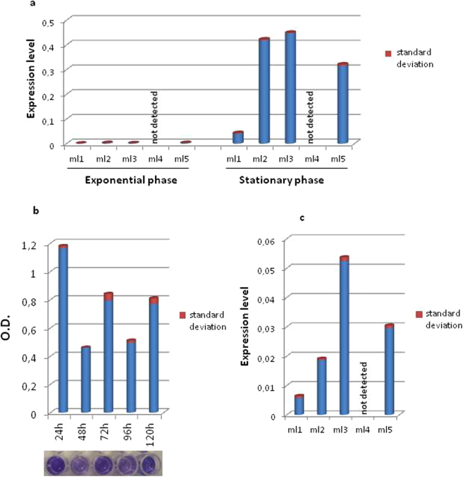
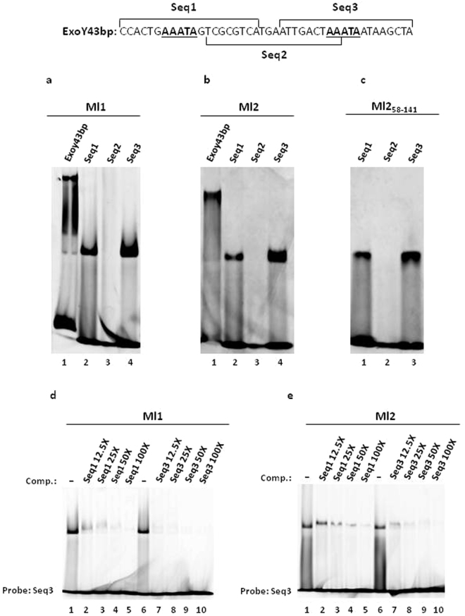
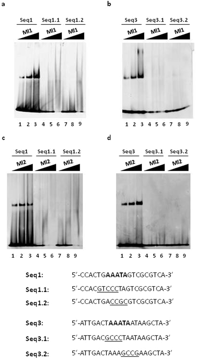
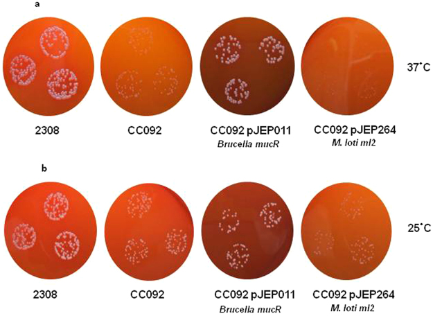
Mesorhizobium loti contains ten genes coding for proteins sharing high amino acid sequence identity with members of the Ros/MucR transcription factor family. Five of these Ros/MucR family members from Mesorhizobium loti (Ml proteins) have been recently structurally and functionally characterized demonstrating that Ml proteins are DNA-binding proteins. However, the DNA-binding studies were performed using the Ros DNA-binding site with the Ml proteins. Currently, there is no evidence as to when the Ml proteins are expressed during the Mesorhizobium lo ti life cycle as well as no information concerning their natural DNA-binding site. In this study, we examine the ml genes expression profile in Mesorhizobium loti and show that ml1, ml2, ml3 and ml5 are expressed during planktonic growth and in biofilms. DNA-binding experiments show that the Ml proteins studied bind a conserved AT-rich site in the promoter region of the exoY gene from Mesorhizobium loti and that the proteins make important contacts with the minor groove of DNA. Moreover, we demonstrate that the Ml proteins studied form higher-order oligomers through their N-terminal region and that the same AT-rich site is recognized by MucR from Brucella abortus using a similar mechanism involving contacts with the minor groove of DNA and oligomerization.


Conflict of interest statement

The authors declare that they have no competing interests.

Figures

Figure 1
Figure 1
Expression profile of ml genes and biofilms formation. (a) ml genes expression level during exponential and planktonic growth; (b) graphic representation of biofilms attachment and detachment phases; (c) ml genes expression level in biofilms at 24 h. For all the RT-qPCR values t-test, P < 0.05.
Figure 2
Figure 2
Mls binding to the Exo43bp, Seq1, Seq2 and Seq3 double-stranded oligonucleotides by EMSA. The nucleotide sequence of the Exo43bp is shown and the sequences of the three 20 bp long oligonucleotides, Seq1, Seq2 and Seq3, are also indicated. (a) EMSA with Ml1; (b) EMSA with Ml2; (c) EMSA with Ml258–141; (d) competition assays with Ml1 using Seq1 and Seq3 as competitors (Comp.) and the FAM-labelled Seq3 as a probe; (e) competition assays with Ml2 using Seq1 and Seq3 as competitors (Comp.) and the FAM-labelled Seq3 as a probe. The amounts of the competitors are indicated on the top of the lanes as excess with respect to the probe (from 12.5-fold to 100-fold excess). Full-length gels are presented in Supplementary Figs S8–S12.
Figure 3
Figure 3
Mls binding to the Seq1 and Seq3 mutant double-stranded oligonucleotides. The sequences of the oligonucleotides tested are shown. The five base pairs shared by Seq1 and Seq3 are in bold and the mutated bases in Seq1.1, Seq1.2, Seq3.1 and Seq3.2 are underlined. Three different quantities of each protein (0.5 μg, 1 μg, and 2 μg) were used with each double-stranded oligonucleotides. The growing quantities of protein used is indicated on the top of each lane (a) EMSA of Ml1 with Seq1, Seq1.1 and Seq1.2; (b) EMSA of Ml1 with Seq3, Seq3.1, Seq3.2; (c) EMSA of Ml2 with Seq1, Seq1.1 and Seq1.2; (d) EMSA of Ml2 with Seq3, Seq3.1, Seq3.2. Full-length gels are presented in Supplementary Figs S13–S16.
Figure 4
Figure 4
Mls binding to the core double-stranded oligonucleotides (ad) and competition experiments (e–l). The sequences of the oligonucelotides tested as target sites and as competitors (Comp.) are shown. The competition experiments were perfomed by adding 25-, 50- or 100-fold excess of each competitor to the reaction mixture with respect to the amount of the FAM-labelled Seq3 double-stranded oligonucleotide used as the probe. Full-length gel are presented in Supplementary Figs S17–S26.
Figure 5
Figure 5
Competition assays of Mls DNA-binding with netropsin. (a) Competition assay with Ml1 binding to double-stranded core2 and (b) to double-stranded core5 in the presence of a 1:1 molar ratio of netropsin; (c) competition assay of Ml2 binding to double-stranded core2 and (d) to double-stranded core5 in the presence of a 1:1 molar ratio of netropsin. Full-length gels are presented in Supplementary Figs S27–S30.
Figure 6
Figure 6
Brucella MucR binding to the target site for the Mls. (a) EMSA of MucR binding to the double-stranded oligonucleotides Exoy43bp, Seq1, Seq2 and Seq3; (b) EMSA of MucR binding to the double-stranded oligonucleotides Seq1.1, Seq1.2, Seq3.1 and Seq3.2; (c) competition assay of MucR binding to double-stranded Seq3 in the presence of a 1:1 molar ratio of netropsin. Full-length gels are presented in Supplementary Figs S31–S33.
Figure 7
Figure 7
Colony formation by B. abortus 2308, an isogenic mucR mutant CC092, CC092 carrying a plasmid-borne copy of the Brucella mucR [CC092 (pJEP011) and CC092 carrying a plasmid-borne copy of the M. loti ml2 under control of the Brucella mucR promoter [CC092 (pJEP264)] following 72 h incubation at 37 °C (a) and 168 h incubation at 25 °C (b) on Schaedler agar supplemented with 5% defibrinated bovine blood. Colony size of the mucR mutant CC092 is restored by transforming the mutant strain with pJEP011 containing the mucR gene, and its native promoter, whereas colony size is not restore by plasmid pJEP264 containing the ml2 gene under control of mucR promoter, under the condition tested.

References

    1. Baglivo I, et al. The structural role of the zinc ion can be dispensable in prokaryotic zinc-finger domains. Proc. Natl. Acad. Sci. US A. 2009;106:6933–6938. doi: 10.1073/pnas.0810003106. - DOI - PMC - PubMed
    1. Caswell CC, et al. Diverse genetic regulon of the virulence-associated transcriptional regulator MucR in Brucella abortus. Infection and Immunity. 2013;81:1040–1051. doi: 10.1128/IAI.01097-12. - DOI - PMC - PubMed
    1. Netti F, et al. An experimentally tested scenario for the structural evolution of eukaryotic Cys2His2 zinc fingers from eubacterial ros homologs. Mol. Biol. Evol. 2013;30:1504–1513. doi: 10.1093/molbev/mst068. - DOI - PubMed
    1. Close TJ, Tait RC, Kado CI. Regulation of Ti plasmid virulence genes by a chromosomal locus of Agrobacterium tumefaciens. J. Bacteriol. 1985;164:774–781. - PMC - PubMed
    1. Mueller K, González JE. Complex regulation of symbiotic functions is coordinated by MucR and quorum sensing in Sinorhizobium meliloti. J. Bacteriol. 2011;193:485–496. doi: 10.1128/JB.01129-10. - DOI - PMC - PubMed

MeSH terms

LinkOut - more resources