. 2017 Nov 17;7(1):15767.
doi: 10.1038/s41598-017-16116-8.

Phosphatidylserine decarboxylase CT699, lysophospholipid acyltransferase CT775, and acyl-ACP synthase CT776 provide membrane lipid diversity to Chlamydia trachomatis

Affiliations

Phosphatidylserine decarboxylase CT699, lysophospholipid acyltransferase CT775, and acyl-ACP synthase CT776 provide membrane lipid diversity to Chlamydia trachomatis

Eric Soupene et al. Sci Rep. .

Abstract

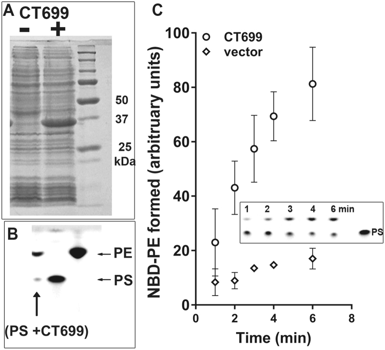
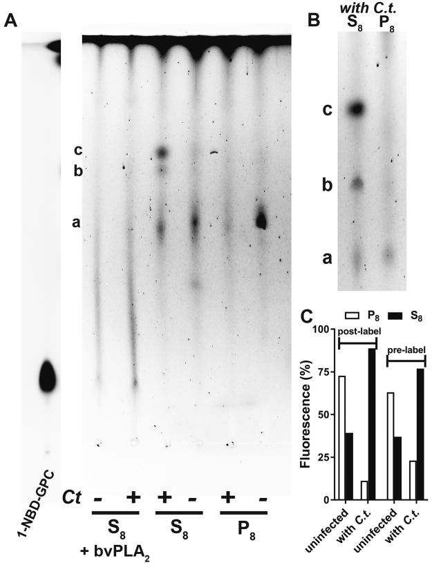
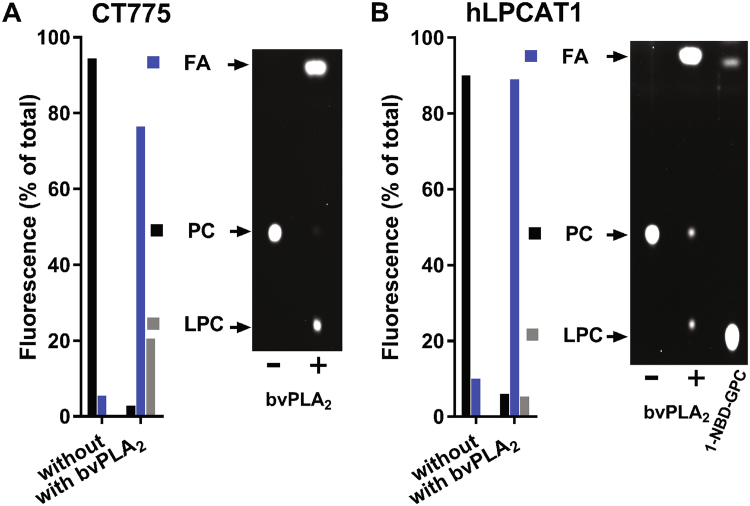
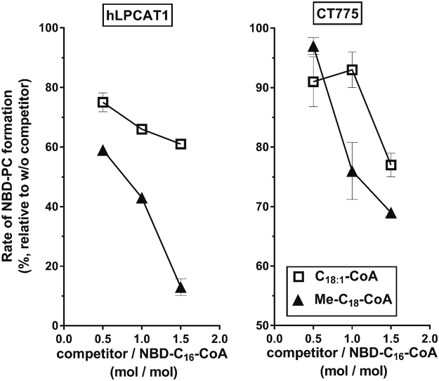
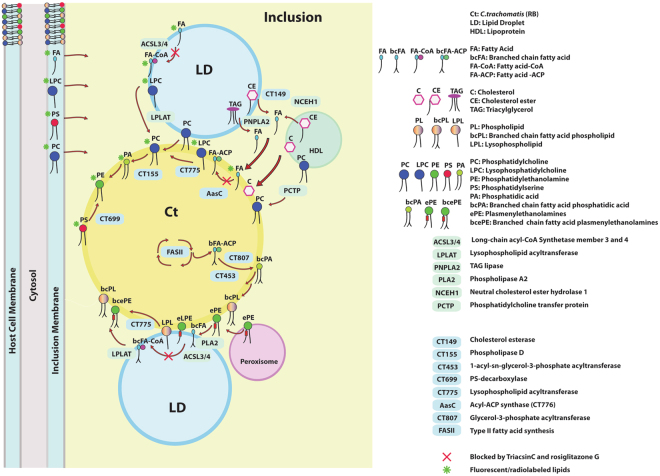
De novo lipid synthesis and scavenging of fatty acids (FA) are processes essential for the formation of the membrane of the human pathogen Chlamydia trachomatis (C.t.). Host FA are assimilated via esterification by the bacterial acyl-acyl carrier protein (ACP) synthase AasC but inhibitors of the host acyl-CoA synthetase enymes ACSL also impaired growth of C.t. in human cells. In E. coli, activity of AasC was sensitive to triacsin C and rosiglitazone G. The absence of a triacsin C-insensitive pathway and the increased inhibition by rosiglitazone G confirmed the sensitivity of the bacterial acyl-ACP synthase to these drugs in infected human cells. We found no evidence that the human ACSL enzymes are required for lipid formation by C.t. The broad substrate specificity of acyltransferase CT775 provides C.t. with the capacity to incorporate straight-chain and bacterial specific branched-chain fatty acids. CT775 accepts both acyl-ACP and acyl-CoA as acyl donors and, 1- or 2-acyl isomers of lysophosphoplipids as acyl acceptors. The enzyme responsible for remodeling of human phosphatidylserine to bacterial phosphatidylethanolamine was identified as CT699. These findings provide evidence that the pathogen has the ability to extend the lipid diversity of its membrane.


Conflict of interest statement

The authors declare that they have no competing interests.

Figures

Figure 1
Figure 1
In vivo labeling of lipids in Chlamydia-infected cells. (A) HeLa cells were grown in T-75 flasks and were infected with C.t. or maintained uninfected. After 24 to 30 hours of infection, the cells were labeled by addition of 5 µM C1-BODIPY500/510 C12 to the medium, After incubation at 37 °C for an additional 2 hours, the cells were washed, and fresh medium containing triacsin C (10 µM), rosiglitazone G (100 µM) or ethanol (untreated cells) was added. After 1 hour of incubation at 37 °C, 10 µM 14C-C16-OH complexed with BSA was added to the medium. The cells were washed and harvested after a further 1-hour incubation at 37 °C (see Methods), and lipids and proteins were extracted. (A) Lipids were separated by thin-layer chromatography, and dually labeled lipids were visualized with a FluoChem camera and after exposure to a phosphorimager screen scanned with a StormImager. A sample obtained from HeLa-infected cells (HeLa/Ct) is shown in lane 1 of the TLC plate. C1-BODIPY500/510 C12 (lane 2) and 14C-C16-OH (lane 3) were used as migration standards. The migration positions of FA and phospholipids are indicated on the left. (B) Proteins were separated by SDS-PAGE, transferred to a PVDF membrane and blotted with monoclonal antibodies against C.t. HSP60 (upper panels) and human GAPDH (lower panel).
Figure 2
Figure 2
Quantification of in vivo FA incorporation in the presence of acyl-CoA synthetase inhibitors. Phospholipids and TAG obtained from dually-labeled HeLa cells infected with C.t. or maintained uninfected were separated by TLC, and fluorescence and radiolabeled lipids were quantified as described in the legend to Fig. 1. The cells were treated with 10 µM triacsin C and 100 µM rosiglitazone G, both of which were dissolved in ethanol. Ethanol was added to the untreated cells at the same final concentration as to the treated cells. The values obtained after labeling of lipids by the fatty acid analog C1-BODIPY500/510 C12 were used to calculate the % inhibition of incorporation of radiolabeled 14C-C16-OH (panels A and C). Values obtained with untreated cells were used to calculate the change in FA incorporation in the presence of the drugs (panels B and D). Note that whereas triacsin C inhibited FA incorporation in both uninfected and infected cells, rosiglitazone G did not reduce the incorporation of FA into glycerophospholipids of uninfected cells. The error bars indicate the standard deviation of 3 measurements of duplicated experiments.
Figure 3
Figure 3
Sensitivity of the C.t. acyl-ACP synthase AasC to acyl-CoA synthetase inhibitors. AasC (CT776) was expressed in E. coli, and the incorporation of 5 µM 14C-C16-OH was determined in crude lysates in the presence of 5 µM triacsin C and 100 µM rosiglitazone G, as indicated. The reactions were performed at 30 °C; the rate of 14C-PL formation was calculated from measurements of 4 samples taken at intervals between 0 and 8 min. The values obtained in the absence of the drugs were used to calculate % inhibition in their presence. (A) E. coli proteins before and after induction of expression of C.t. AasC were separated by SDS-PAGE and stained with the GelCode Blue dye. Panels B and C. Quantification of the incorporation of 14C-C16-OH in the presence of triacsin C (panel B) and rosiglitazone G (panel C) is reported as percentage relative to the incorporation observed in their absence. Three independent experiments were performed and the error bars indicate the standard deviation of 3 measurements.
Figure 4
Figure 4
Labeling of Chlamydia-infected cells with NBD-PS. HeLa cells were grown on coverslips and infected with C. trachomatis strain D. After 24 hours, 1 µM NBD-PS (green) was added to the medium. The cells were washed and fixed after 1 hour of incubation. DNA was stained with Hoechst dye (blue), and imaging was performed with a Keyence microscope equipped with a 40x objective. Panel B shows a magnified cropped image of an infected cell with 2 inclusions, which are indicated. Images were taken from a single labeling experiment.
Figure 5
Figure 5
CT699 is a PS-decarboxylase enzyme. CT699 was expressed in E. coli (panel A), and activity measurements were performed in the presence of 2 µM NBD-PS at 37 °C for 20 min (panel B, lane PS + CT699) or from 0 to 6 minutes (panel C). Control reactions were performed using E. coli cells without the cloned CT699 construct (vector, panel C). Lipids were extracted and separated by thin-layer chromatography using unreacted NBD-PS and NBD-PE as migration standards (panel B and insert panel C). Several independent experiments were performed and the error bars in panel C indicate the standard deviation of 3 measurements of the samples obtained in one experiment.
Figure 6
Figure 6
Labeling of Chlamydia-infected cells with 1-NBD-GPC. HeLa cells were grown in T-75 flasks and labeled with 5 µM 1-NBD-GPC (green) for 2 hours at 37 °C. The cells were washed and incubated in fresh medium for 2 hours before infection with C. trachomatis strain D. Live imaging was performed after 36 hours using a Keyence microscope equipped with a 20x objective (set a; bright-field and merged images with NBD fluorescence are shown). Note the presence of labeled and unlabeled cells. Cropped images (bright-field, NBD fluorescence, and merged images) showing 2 infected cells with labeled inclusions and one cell with an unlabeled inclusion are shown on the right (set b). Lipids were extracted from the treated cells and analyzed in the experiments presented in Fig. 7. Labeling experiments were performed twice and shown images were taken during the second experiment.
Figure 7
Figure 7
In vivo acylation of 1-NBD-GPC in Chlamydia-infected cells. Cells were grown and labeled as described in the legend of Fig. 6. Cells were either infected after labeling of the cells (pre-labeling conditions; images shown in Fig. 6) or were infected before addition of 1-NBD-GPC (post-labeling conditions; images shown in Figure S2). Cells were collected, washed, and lysed. The lysates were centrifuged at 8,000 g to generate pellet (P8) and supernatant (S8) fractions that represented the organelle and the cytosolic fractions enriched in EBs/RBs of infected cells, respectively. Lipids were extracted from the fractions obtained from pre-labeled cells (panel A) and post-labeled cells (panel B) and separated by TLC. Fluorescence quantification is shown in panel C. Labeled lipids obtained from pre-labeled cells were treated with bee venom PLA2 and analyzed (S8 + bvPLA2 in panel A). 1-NBD-GPC, which was used as a migration standard, was run on the TLC plate shown on panel A. The fluorescence of the lipids obtained from the samples was weaker than that of the standards, and the gamma intensity of the standard lane was adjusted to match that of the samples. For clarity, the image manipulation is indicated by presenting the standard lane as a separate section of the TLC plate on the left (NBD-LPC lane).
Figure 8
Figure 8
Acyl-CoA::1-acyl-GPC acyltransferase activity of CT775. CT775 was produced in E. coli, and activity in the membrane fraction was measured as described previously. Reactions were performed with 1 µM NBD-C16-CoA in the presence of 10 µM synthetic 1-C16-sn-glycerol-3-P-choline (>90% 1-acyl-GPC isomer) at 30 °C. NBD-PC obtained after 30 minutes reaction with 40 µg of CT775 microsomes was extracted and separated by TLC. The NBD-PC spot was scraped from the plate and treated with bee venom PLA2 (bvPLA2). Lipids were extracted and separated by TLC, and the fluorescence in LPC (1- or 2-acyl-GPC), FA and unreacted PC was quantified. Experiments were performed with CT775 (panel A) and human LPCAT1 (panel B). NBD-LPC was used as a migration standard; it is shown in the right-hand lane of the TLC plate in panel B.
Figure 9
Figure 9
Odd-chain acyl-CoAs are substrates of hLPCAT1 and CT775. Human long-chain acyl-CoA synthetase 6 (hACSL6) was purified and used to produce CoA in the ester form from 17-methyl-stearic acid (MeC18-OH). MeC18-CoA was purified on a Lipidex-1000 column (see Methods), and its ability to compete with the acylation of 1-acyl-GPC from NBD-C16-CoA by human LPCAT1 (panel A) and CT775 (panel B) was determined. Measurements were performed with 0.5 µg of hLPCAT1 or CT775 microsomes in the presence of 20 µM 1-C16-GPC and 0.5 µM NBD-C16-CoA. The competitors C18:1-CoA and MeC18-CoA were added at concentrations of 0.25, 0.5 and 1.0 µM. For each condition, the rate of formation of NBD-PC was calculated from measurements of 3 samples taken between 0 and 6 min. The values obtained in the presence of the competitors are reported as percentages of the values obtained in their absence. Synthesis of odd-chain acyl-CoA was performed twice and were used in several independent competition experiments. The error bars indicate the standard deviation of 3 measurements.
Figure 10
Figure 10
Model for FA and glycerophospholipids scavenging in Chlamydia-infected human cells. The inclusion contains pathways supporting the scavenging and remodeling of lipids obtained from the host membranes, organelles and vesicles. The model only presents enzymes and reactions that relate to the findings presented in this work, and that are described in detail in the Discussion section. Pathways essential for lipid de novo synthesis are not depicted and several pathways are simplified for clarity. Proposed mechanisms for the labeling of Chlamydia lipids by exogenously added fluorescent and radiolabeled lipids (PC, PS, LPC and FA) are highlighted with a green asterisk. Reactions that are sensitive to inhibition by the drugs triacsin C and rosiglitazone G are indicated with a red X.

Similar articles

Cited by

References

    1. Hackstadt T, Fischer ER, Scidmore MA, Rockey DD, Heinzen RA. Origins and functions of the chlamydial inclusion. Trends Microbiol. 1997;5:288–293. doi: 10.1016/S0966-842X(97)01061-5. - DOI - PubMed
    1. Omsland A, Sixt BS, Horn M, Hackstadt T. Chlamydial metabolism revisited: interspecies metabolic variability and developmental stage-specific physiologic activities. FEMS Microbiol. Rev. 2014;38:779–801. doi: 10.1111/1574-6976.12059. - DOI - PMC - PubMed
    1. Boncompain G, et al. The intracellular bacteria Chlamydia hijack peroxisomes and utilize their enzymatic capacity to produce bacteria-specific phospholipids. PLoS One. 2014;9:e86196. doi: 10.1371/journal.pone.0086196. - DOI - PMC - PubMed
    1. Cox JV, Abdelrahman YM, Peters J, Naher N, Belland RJ. Chlamydia trachomatis utilizes the mammalian CLA1 lipid transporter to acquire host phosphatidylcholine essential for growth. Cell Microbiol. 2016;18:305–318. doi: 10.1111/cmi.12523. - DOI - PubMed
    1. Wylie JL, Hatch GM, McClarty G. Host cell phospholipids are trafficked to and then modified by Chlamydia trachomatis. J. Bacteriol. 1997;179:7233–7242. doi: 10.1128/jb.179.23.7233-7242.1997. - DOI - PMC - PubMed

MeSH terms

LinkOut - more resources