. 2018 Jun;175(12):2272-2283.
doi: 10.1111/bph.14098. Epub 2017 Dec 29.

GI-530159, a novel, selective, mechanosensitive two-pore-domain potassium (K2P ) channel opener, reduces rat dorsal root ganglion neuron excitability

Affiliations

GI-530159, a novel, selective, mechanosensitive two-pore-domain potassium (K2P ) channel opener, reduces rat dorsal root ganglion neuron excitability

Alexandre J C Loucif et al. Br J Pharmacol. 2018 Jun.

Abstract

Background and purpose: TREK two-pore-domain potassium (K2P ) channels play a critical role in regulating the excitability of somatosensory nociceptive neurons and are important mediators of pain perception. An understanding of the roles of TREK channels in pain perception and, indeed, in other pathophysiological conditions, has been severely hampered by the lack of potent and/or selective activators and inhibitors. In this study, we describe a new, selective opener of TREK channels, GI-530159.

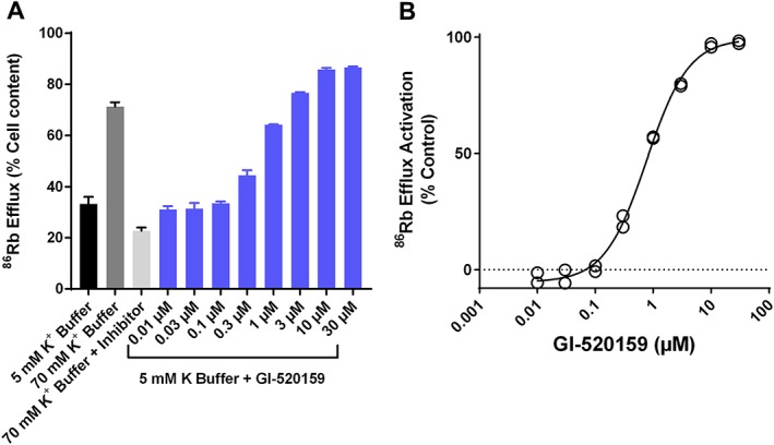
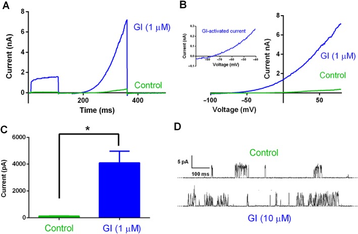
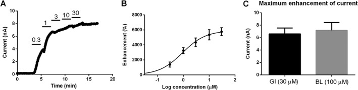
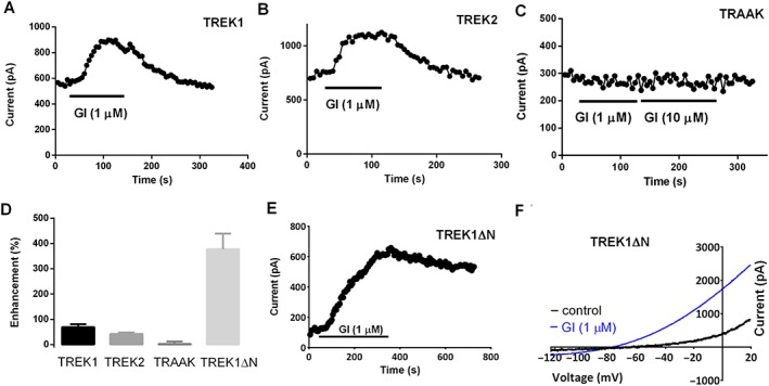
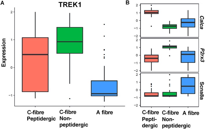
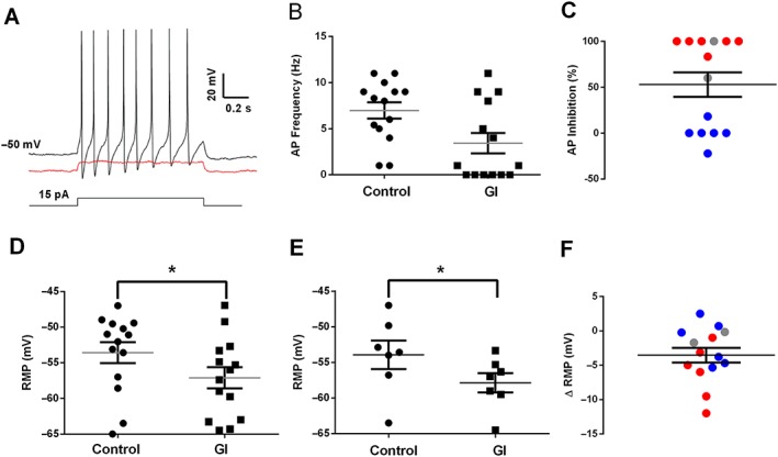
Experimental approach: The effect of GI-530159 on TREK channels was demonstrated using 86 Rb efflux assays, whole-cell and single-channel patch-clamp recordings from recombinant TREK channels. The expression of K2P 2.1 (TREK1), K2P 10.1 (TREK2) and K2P 4.1 (TRAAK) channels was determined using transcriptome analysis from single dorsal root ganglion (DRG) cells. Current-clamp recordings from cultured rat DRG neurons were used to measure the effect of GI-530159 on neuronal excitability.

Key results: For recombinant human TREK1 channels, GI-530159 had similar low EC50 values in Rb efflux experiments and electrophysiological recordings. It activated TREK2 channels, but it had no detectable action on TRAAK channels nor any significant effect on other K channels tested. Current-clamp recordings from cultured rat DRG neurones showed that application of GI-530159 at 1 μM resulted in a significant reduction in firing frequency and a small hyperpolarization of resting membrane potential.

Conclusions and implications: This study provides pharmacological evidence for the presence of mechanosensitive TREK K2P channels in sensory neurones and suggests that development of selective K2P channel openers like GI-530159 could aid in the development of novel analgesic agents.

Linked articles: This article is part of a themed section on Recent Advances in Targeting Ion Channels to Treat Chronic Pain. To view the other articles in this section visit http://onlinelibrary.wiley.com/doi/10.1111/bph.v175.12/issuetoc.


Figures

Figure 1
Figure 1
Structure of GI‐530159.
Figure 2
Figure 2
Effect of GI‐530159 on 86Rb flux through TREK1 channels. (A) Exemplar values of 86Rb efflux from 96‐well plate containing monolayers of CHO–hTREK1 during 60 min exposure to normal 5 mM K EBSS buffer, 70 mM K buffer, 70 mM KCl buffer and reference inhibitor (100 μM CP‐338818) as well increasing concentrations of GI‐530159 made up in 5 mM K EBSS buffer. 86Rb efflux is expressed as % of cell content at beginning of experiment (n = 3 for each concentration of GI‐530159, n = 24 for 5 mM K buffer). (B) Concentration‐dependence of stimulation of 86Rb efflux by GI‐530159 normalized to the response observed with 70 mM K EBSS buffer. Maximal flux (142 ± 2%) and EC50 (0.76 ± 0.1 μM) were derived from fit of logistic equation to data.
Figure 3
Figure 3
Effect of GI‐530159 on stably transfected TREK1 channel currents. (A) Whole‐cell current recordings of TREK1 channels stably transfected in HEK293 cells, in the presence (blue) and absence (green) of GI‐530159 (1 μM). (B) Current–voltage relationship for TREK1 currents in the presence (blue) and absence of GI‐530159. The inset shows the current–voltage relationship for GI‐530159‐activated current. (C) Control current, measured at 0 mV (green), was significantly enhanced by 1 μM GI‐530139 (blue, n = 6, *P < 0.05, paired t‐test). Each n value represents a recording from a cell on an independent coverslip on different recording days. (D) Representative single‐channel records of hTREK‐1 in excised inside‐out membrane patches (12 inside‐out patch recordings in total) from HEK293 cells in the presence and absence of GI‐530159 (10 μM). Dotted line indicates the closed channel state, and upward deflections correspond to channel openings. Membrane patches were voltage clamped at +60 mV at room temperature.
Figure 4
Figure 4
Concentration–response curve for GI‐530159 on stably transfected TREK1 channels. (A) Representative example of cumulative concentration–response curve for GI‐530159 on TREK1 current stably transfected in HEK293 cells. Current was measured at 0 mV. (B) Concentration–response curve reveals EC50 of 0.9 μM for GI‐530159 (n = 6 for each concentration). Each n value represents a recording from a cell on an independent coverslip on different recording days. (C) Maximum current enhancement by GI‐530159 (n = 6) is similar to that seen for BL‐1249 (n = 7) for TREK1 currents in stably transfected HEK293 cells.
Figure 5
Figure 5
Effect of GI‐530159 on TREK1, TREK2, TRAAK and TREK1ΔN channels transiently transfected in tsA‐201 cells. (A, B) GI‐530159 activates TREK1 and TREK2 channels transiently transfected in tsA‐201 cells. (C) GI‐530159 has no detectable activation of TRAAK channels. (D) Effect of GI‐530159 on TREK1 (n = 6), TREK2 (n = 6), TRAAK (n = 7) and TREK1ΔN (n = 5) channels in transiently transfected tsA‐201 cells. (E, F) GI‐530159 activates TREK1ΔN channels. Each n value represents a recording from a cell on an independent coverslip on different recording days. The degree of enhancement of current through TREK1 channels was found to be not significantly different from that through TREK2 channels but was significantly smaller than that through TREK1ΔN channels (one‐way ANOVA, followed by Dunnett's multiple comparisons test; P<0.05).
Figure 6
Figure 6
Single DRG neuron transcriptome – TREK1 channels. (A) Differential TREK1 expression in single peptidergic C fibres, non‐peptidergic C fibres and A fibres. (B) Comparative expression of selective markers for peptidergic C fibres (Calca), non‐peptidergic C fibres (P2rx3) and A fibres (Scn8a). Expression is given as log2 FPKM. Data are from 120 individual DRG neurons isolated from four rats.
Figure 7
Figure 7
Effect of GI‐530159 on small DRG neuron firing properties. (A) Normal firing of individual DRG neuron in response to current injection (black trace) is inhibited by GI‐530159 (1 μM, red trace). (B, C) GI‐530159 inhibits action potential firing in some small DRG neurons. (D–F) GI‐530159 (1 μM) hyperpolarizes the membrane potential of small DRG neurons. In (D), membrane potential in each individual neuron is hyperpolarized from −53.6 ± 1.5 to −57.1 ± 1.5 mV (n = 14 individual neurons, P < 0.05, paired t‐test) by GI‐530159 (1 μM). In (E), the average membrane potential in neurons from a given rat is significantly hyperpolarized from −54.0 ± 2.0 to −57.8 ± 1.4 mV (n = 7 rats, P < 0.05, paired t‐test, Figure 7D) by GI‐530159 (1 μM). In (C, F), six neurons with clear inhibition of firing are shown in red, and six neurons where there was no inhibition of firing are in blue. Two neurons, which were not clearly defined, are shown in grey. Each recorded neuron was from an independent coverslip, and the 14 neurons were taken from seven independent preparations of DRG neurons.

References

    1. Acosta C, Djouhri L, Watkins R, Berry C, Bromage K, Lawson SN (2014). TREK2 expressed selectively in IB4‐binding C‐fiber nociceptors hyperpolarizes their membrane potentials and limits spontaneous pain. J Neurosci 34: 1494–1509. - PMC - PubMed
    1. Alexander SPH, Peters JA, Kelly E, Marrion NV, Faccenda E, Harding SD et al (2017a). The Concise Guide to PHARMACOLOGY 2017/18: Ligand‐gated ion channels. Br J Pharmacol 174: S130–S159. - PMC - PubMed
    1. Alexander SPH, Striessnig J, Kelly E, Marrion NV, Peters JA, Faccenda E et al (2017b). The Concise Guide to PHARMACOLOGY 2017/18: Voltage‐gated ion channels. Br J Pharmacol 174: S160–S194. - PMC - PubMed
    1. Alloui A, Zimmermann K, Mamet J, Duprat F, Noël J, Chemin J et al (2006). TREK1, a K+ channel involved in polymodal pain perception. EMBO J 25: 2368–2376. - PMC - PubMed
    1. Bagriantsev SN, Ang KH, Gallardo‐Godoy A, Clark KA, Arkin MR, Renslo AR et al (2013). A high‐throughput functional screen identifies small molecule regulators of temperature‐ and mechano‐sensitive K2P channels. ACS Chem Biol 8: 1841–1851. - PMC - PubMed

Publication types

Substances

LinkOut - more resources