

Thalamic volume reduction in drug-naive patients with new-onset genetic generalized epilepsy
- PMID: 29150855
- PMCID: PMC5813228
- DOI: 10.1111/epi.13955
Thalamic volume reduction in drug-naive patients with new-onset genetic generalized epilepsy
Abstract
Objective: Patients with genetic generalized epilepsy (GGE) have subtle morphologic abnormalities of the brain revealed with magnetic resonance imaging (MRI), particularly in the thalamus. However, it is unclear whether morphologic abnormalities of the brain in GGE are a consequence of repeated seizures over the duration of the disease, or are a consequence of treatment with antiepileptic drugs (AEDs), or are independent of these factors. Therefore, we measured brain morphometry in a cohort of AED-naive patients with GGE at disease onset. We hypothesize that drug-naive patients at disease onset have gray matter changes compared to age-matched healthy controls.
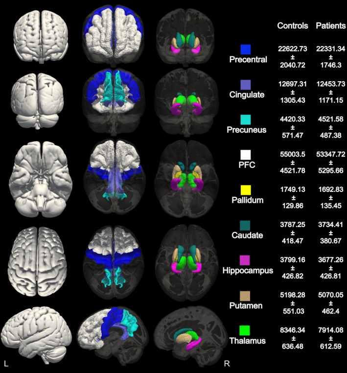
Methods: We performed quantitative measures of gray matter volume in the thalamus, putamen, caudate, pallidum, hippocampus, precuneus, prefrontal cortex, precentral cortex, and cingulate in 29 AED-naive patients with new-onset GGE and compared them to 32 age-matched healthy controls. We subsequently compared the shape of any brain structures found to differ in gray matter volume between the groups.
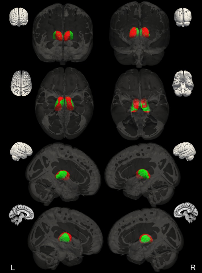
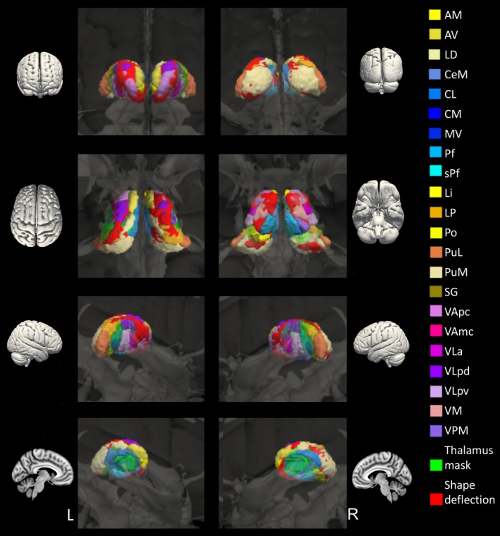
Results: The thalamus was the only structure to show reduced gray matter volume in AED-naive patients with new-onset GGE compared to healthy controls. Shape analysis revealed that the thalamus showed deflation, which was not uniformly distributed, but particularly affected a circumferential strip involving anterior, superior, posterior, and inferior regions with sparing of medial and lateral regions.
Significance: Structural abnormalities in the thalamus are present at the initial onset of GGE in AED-naive patients, suggesting that thalamic structural abnormality is an intrinsic feature of GGE and not a consequence of AEDs or disease duration.
Keywords: drug naive; genetic generalized epilepsy; new onset; thalamus; volumetric MRI.
© 2017 The Authors. Epilepsia published by Wiley Periodicals, Inc. on behalf of International League Against Epilepsy.
Figures
Comment in
-
Brain morphological abnormalities in genetic generalized epilepsies: The starting point?Epilepsia. 2019 Jul;60(7):1279-1280. doi: 10.1111/epi.16101. Epub 2019 Jun 24. Epilepsia. 2019. PMID: 31233212 No abstract available.
References
-
- Berg AT, Berkovic SF, Brodie MJ, et al. Revised terminology and concepts for organization of seizures and epilepsies: report of the ILAE Commission on Classification and Terminology, 2005‐2009. Epilepsia. 2010;51:676–85. - PubMed
-
- Du H, Zhang Y, Xie B, et al. Regional atrophy of the basal ganglia and thalamus in idiopathic generalized epilepsy. J Magn Reson Imaging. 2011;33:817–21. - PubMed
-
- Seeck M, Dreifuss S, Lantz G, et al. Subcortical nuclei volumetry in idiopathic generalized epilepsy. Epilepsia. 2005;46:1642–5. - PubMed
-
- Lin K, de Araujo Filho GM, Pascalicchio TF, et al. Hippocampal atrophy and memory dysfunction in patients with juvenile myoclonic epilepsy. Epilepsy Behav. 2013;29:247–51. - PubMed
Publication types
MeSH terms
Grants and funding
LinkOut - more resources
Full Text Sources
Other Literature Sources
