. 2018 Jan;33(1):159-164.
doi: 10.1002/mds.27215. Epub 2017 Nov 18.

Directional local field potentials: A tool to optimize deep brain stimulation

Affiliations

Directional local field potentials: A tool to optimize deep brain stimulation

Gerd Tinkhauser et al. Mov Disord. 2018 Jan.

Abstract

Background: Although recently introduced directional DBS leads provide control of the stimulation field, programing is time-consuming.

Objectives: Here, we validate local field potentials recorded from directional contacts as a predictor of the most efficient contacts for stimulation in patients with PD.

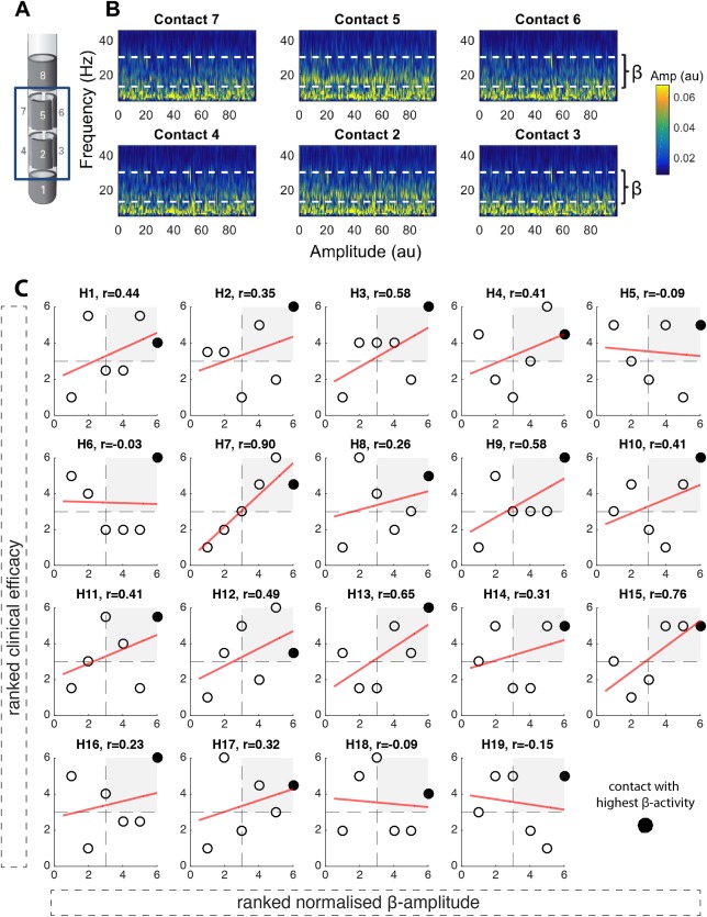
Methods: Intraoperative local field potentials were recorded from directional contacts in the STN of 12 patients and beta activity compared with the results of the clinical contact review performed after 4 to 7 months.

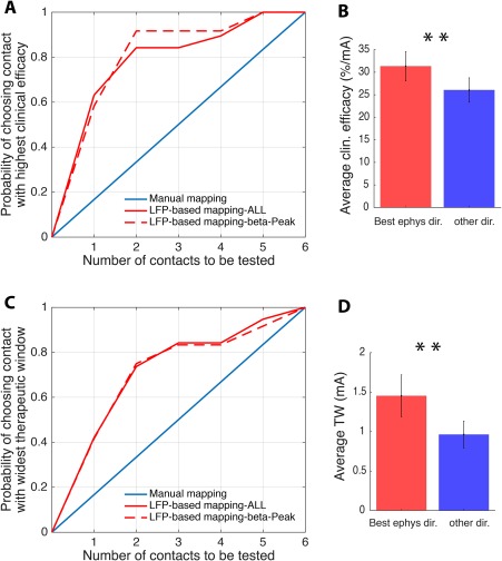
Results: Normalized beta activity was positively correlated with the contact's clinical efficacy. The two contacts with the highest beta activity included the most efficient stimulation contact in up to 92% and that with the widest therapeutic window in 74% of cases.

Conclusion: Local field potentials predict the most efficient stimulation contacts and may provide a useful tool to expedite the selection of the optimal contact for directional DBS. © 2017 The Authors. Movement Disorders published by Wiley Periodicals, Inc. on behalf of International Parkinson and Movement Disorder Society.

Keywords: DBS programming; Parkinson's disease; deep brain stimulation; directional leads; local field potentials.


Figures

Figure 1
Figure 1
Directional LFPs and relationship between ranked beta activity and clinical efficacy. (A) illustrates the directional DBS lead (Boston Scientific, Marlborough, MA). Contacts are distributed along four levels. On levels two and three, there are three segmented contacts (level two: contacts 2/3/4; level three: contacts 5/6/7). (B) shows an example time frequency spectrum from an intraoperative LFP recording (duration, 100 seconds) from the six directional contacts (2/3/4; 5/6/7) with the patient awake and at rest. The dashed white line marks the beta frequency band (13‐35 Hz). It shows that LFP beta activity is not equally distributed across directional contacts. Contact 5 shows the highest beta activity, followed by contact 2, with both contacts 5 and 2 oriented in the same direction. Data from the right hemisphere in subject 3 (for raw data, amplitude‐frequency spectrum, and imaging from the same subject and hemisphere, see Supplementary Fig. 1). (C) illustrates the relationships between normalized beta activity and clinical efficacy across the six directional contacts in each hemisphere (H = hemisphere; n = 19). The normalized beta amplitude is shown on the x‐axis, the clinical efficacy on the y‐axis, and Spearman correlation coefficients are shown on the top of each panel. The best electrophysiological contact (contact with highest normalized beta activity) is highlighted in black. The red linear regression fit is shown only for illustration purposes. In 15 hemispheres, a positive relationship between clinical efficacy and normalized beta activity was found (t 18 = 4.65; P < 0.001, one‐sample t test). In 12 of 19 hemispheres, the contact with the highest beta activity matched the clinically most effective stimulation contact. Furthermore, in all hemispheres, the contact with the highest beta activity was localized in the upper‐right quadrant, where the clinically more efficient contacts are localized. Clinical efficacy and normalized beta activity are illustrated as ranked values; Supplementary Figure 3 shows the same figure with nonranked values. [Color figure can be viewed at wileyonlinelibrary.com]
Figure 2
Figure 2
LFP‐based DBS programming. (A) shows the probability of identifying the stimulation contact with the highest clinical efficacy, comparing the conventional (random) test strategy in blue with the LFP‐based test strategy in red (full red line: all hemispheres n = 19, dashed red line: only hemispheres with clear beta peak n = 12). While for conventional mapping the probability of identifying the most efficient stimulation contact increases by 0.17 with each contact tested, the LFP‐based strategy identifies the most efficient contact with a probability of 0.63 if only the contact with the highest beta activity is considered, and with a probability of 0.84 if the two contacts with the highest beta activity are considered. By considering hemispheres with a clear beta peak only, the probability increases up to 0.92 when the two best electrophysiological contacts are considered. (B) The mean clinical efficacy of the two directional stimulation contacts with the highest beta activity (“Best ephys”) is significantly higher compared to the clinical efficacy of the remaining directional contacts (“other dir.”). (C) (similar to A) shows the probability of identifying the contact with the highest therapeutic window by again comparing the conventional (random) test strategy in blue with the LFP‐based test strategy in red (full red line: all hemispheres n = 19; dashed red line: only hemispheres with clear beta peak n = 12). While for conventional mapping the probability of identifying the stimulation contact with the widest therapeutic window increases by 0.17 with each contact tested, the LFP‐based strategy identifies the contact with the widest therapeutic window with a probability of 0.42 if only the contact with the highest beta activity is considered, and with a probability of 0.74 if the two contacts with the highest beta activity are considered. No relevant difference in the predictive value is found when exclusively hemispheres with a clear beta peak are considered (dashed red line). (D) The mean TW of the two directional stimulation contacts with the highest beta activity (“Best ephys”) is significantly higher compared to the therapeutic window of the remaining directional contacts. Values are mean ± SEM; **P < 0.01. [Color figure can be viewed at wileyonlinelibrary.com]

Comment in

  • Next generation programming.
    Israel Z, Bergman H, Eitan R. Israel Z, et al. Mov Disord. 2018 Feb;33(2):186. doi: 10.1002/mds.27322. Epub 2018 Jan 25. Mov Disord. 2018. PMID: 29369418 No abstract available.

References

    1. Pollo C, Kaelin‐Lang A, Oertel MF, et al. Directional deep brain stimulation: an intraoperative double‐blind pilot study. Brain 2014;137:2015‐2026. - PubMed
    1. Timmermann L, Jain R, Chen L, et al. Multiple‐source current steering in subthalamic nucleus deep brain stimulation for Parkinson's disease (the VANTAGE study): a non‐randomised, prospective, multicentre, open‐label study. Lancet Neurol 2015;14:693‐701. - PubMed
    1. Contarino MF, Bour LJ, Verhagen R, et al. Directional steering: a novel approach to deep brain stimulation. Neurology 2014;83:1163‐1169. - PubMed
    1. Steigerwald F, Müller L, Johannes S, Matthies C, Volkmann J. Directional deep brain stimulation of the subthalamic nucleus: a pilot study using a novel neurostimulation device. Mov Disord 2016;31: 1240‐1243. - PMC - PubMed
    1. Reker P, Dembek TA, Becker J, et al. Directional deep brain stimulation: a case of avoiding dysarthria with bipolar directional current steering. Parkinsonism Relat Disord 2016;31:156‐158. - PubMed

Publication types