Review
. 2018 Jan;34(1):60-68.
doi: 10.1111/phpp.12368.

Drug delivery strategies for chemoprevention of UVB-induced skin cancer: A review

Affiliations
Review

Drug delivery strategies for chemoprevention of UVB-induced skin cancer: A review

Arvind Bagde et al. Photodermatol Photoimmunol Photomed. 2018 Jan.

Abstract

Annually, more skin cancer cases are diagnosed than the collective incidence of the colon, lung, breast, and prostate cancer. Persistent contact with sunlight is a primary cause for all the skin malignancies. UVB radiation induces reactive oxygen species (ROS) production in the skin which eventually leads to DNA damage and mutation. Various delivery approaches for the skin cancer treatment/prevention have been evolving and are directed toward improvements in terms of delivery modes, therapeutic agents, and site-specificity of therapeutics delivery. The effective chemoprevention activity achieved is based on the efficiency of the delivery system used and the amount of the therapeutic molecule deposited in the skin. In this article, we have discussed different studies performed specifically for the chemoprevention of UVB-induced skin cancer. Ultra-flexible nanocarriers, transethosomes nanocarriers, silica nanoparticles, silver nanoparticles, nanocapsule suspensions, microemulsion, nanoemulsion, and polymeric nanoparticles which have been used so far to deliver the desired drug molecule for preventing the UVB-induced skin cancer.

Keywords: UVB radiation; chemoprevention; drug delivery strategy; skin cancer.


Figures

FIGURE 1
FIGURE 1
Anatomy of skin showing types of skin cells. Reproduced from Korotkov K et al.
FIGURE 2
FIGURE 2
Electromagnetic spectrum of visible and UV radiation and its biologic effects on human skin. UVA generates reactive oxygen species which causes DNA damage through photosensitizing reactions. UVB-radiation-induced DNA damage leads to molecular re-arrangements which eventually results in formation of 6-4 photoproducts and cyclobutane dimers. Reproduced from D’Orazio J et al
FIGURE 3
FIGURE 3
A, DIM-D deposition in the skin at the end of 24 h from PEG solution, NLC, NLC-OA, and UltraFLEX-Nano. B, Drug released from 0.3, 1.0, and 1.5% DIM-D-UltraFLEX-Nano HPMC gels. Data has been represented as the mean ± SD. ***P < .001. Reproduced from Boakye CH et al.
FIGURE 4
FIGURE 4
A, Tumors formed in UV-only-irradiated group and DIM-D-UltraFLEX-Nano-gel-treated group. B, Percentage of animals showing tumors at the onset of tumorigenesis. Reproduced from Boakye CH et al.
FIGURE 5
FIGURE 5
Drug permeation through skin from NPZ-DM and NPZ-TE formulations. Values are expressed as mean ± SD (n = 3). Statistical analysis: *P < .05 vs NPZ-H2 and NPZ-TE, **P < .001 vs NPZ-H2O, NPZ-TE, and NPZ-DM. Reproduced from Menezes AC, et al.
FIGURE 6
FIGURE 6
Skin retention after 24 h of free Quercetin (black colored) and Q/NH2-MSN_1/1 (gray colored) from different media. Each bar represents the mean ± SD obtained in three independent experiments. Reproduced from Sapino S et al.
FIGURE 7
FIGURE 7
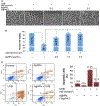
Decrease in apoptosis and UVB-induced cell death on pretreatment with silver nanoparticles. HaCaT cells (1 Å~ 106/plate) were grown in UVtransparent glass plates and treated with AgNPs 3 h prior to UVB exposure. After 24-h exposure of UVB radiation (A) images of the cells under phase-contrast microscope. Cells not irradiated with UVB were used as a control. Arrows indicate apoptotic cells. B, Percent cell viability in different treatment groups. Bars represent mean ± SD, n = 3; *P < .05; **P < .01. C Early apoptotic cells percent in each treatment group. Bars represent mean ± SD, n = 3; **P < .01. Reproduced from Arora S et al.
FIGURE 8
FIGURE 8
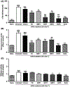
Anti-inflammatory effects of semisolid formulations on UVB-irradiation-induced skin injury in mice. Ear edema (A), myeloperoxidase (B), and NAGase (C) activities in mice submitted to UVB irradiation (0.5 J/cm2). All formulations (15 mg/ear) were immediately applied after UVB irradiation. Ear edema and cell infiltration were measured 24 h after irradiation. Each bar represents the mean + SEM (n = 6-7); ***P < .001 when compared to the naïve group. *P < .05, **P < .01, and ***P < .001 when compared to the untreated group. & P < .01 shows significant difference between semisolids containing Q10-loaded nanocapsules (NCP1 and NCP3) with semisolids containing nanocapsules without Q10 (NBP1 and NBP3). One-way ANOVA followed by post hoc Newman-Keuls test. Reproduced from Pegoraro NS et al.
FIGURE 9
FIGURE 9
Results of apoptotic sunburn cells quantitation in epidermis of normal skin, skin treated with UVB, skin treated with UVB+Vehicle, and skin treated with UVB+CAF. Values are mean ± SD (n = 3). *P < .05. Reproduced from Ma H et al.

References

    1. Boakye CH, Patel K, Doddapaneni R, et al. Ultra-flexible nanocarriers for enhanced topical delivery of a highly lipophilic antiaxidative molecule for skin cancer chemoprevention. Colloids Surf B. 2016;143:156–167. - PMC - PubMed
    1. D’Orazio J, Jarrett S, Amaro-Ortiz A, Scott T. UV radiation and the skin. Int J Mol Sci. 2013;14:12222–12248. - PMC - PubMed
    1. Simões M, Sousa J, Pais A. Skin cancer and new treatment perspectives: A review. Cancer Lett. 2015;357:8–42. - PubMed
    1. Armstrong BK, Kricker A. The epidemiology of UV induced skin cancer. J Photochem Photobiol, B. 2001;63:8–18. - PubMed
    1. Narayanan DL, Saladi RN, Fox JL. Ultraviolet radiation and skin cancer. Int J Dermatol. 2010;49:978–986. - PubMed