

Isolated Pancreatic Myeloid Sarcoma Associated with t(8;21)/RUNX1-RUNX1T1 Rearrangement
- PMID: 29151502
- PMCID: PMC5849554
- DOI: 10.2169/internalmedicine.8912-17
Isolated Pancreatic Myeloid Sarcoma Associated with t(8;21)/RUNX1-RUNX1T1 Rearrangement
Abstract
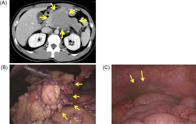
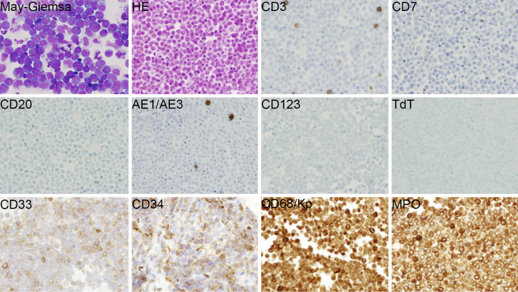
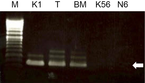
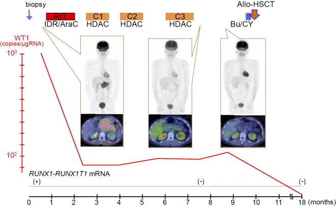
No valid treatment for isolated myeloid sarcoma (IMS) has yet been established, and no thorough genetic examinations have been performed because of its low incidence and unique manner of development. We herein report a 34-year-old man with pancreatic IMS with t(8;21)/RUNX1-RUNX1T1 rearrangement. He was treated with high-dose cytarabine followed by allogeneic hematopoietic stem cell transplantation (allo-HSCT). This is the first report of pancreatic IMS with t(8;21). Positron emission tomography/computed tomography and genetic study are useful for the diagnosis, and allo-HSCT achieved complete remission in this patient.
Keywords: isolated myeloid sarcoma; pancreas; t(8;21).
Figures
Similar articles
-
The dynamics of RUNX1-RUNX1T1 transcript levels after allogeneic hematopoietic stem cell transplantation predict relapse in patients with t(8;21) acute myeloid leukemia.J Hematol Oncol. 2017 Feb 6;10(1):44. doi: 10.1186/s13045-017-0414-2. J Hematol Oncol. 2017. PMID: 28166825 Free PMC article.
-
Myeloid neoplasms with t(16;21)(q24;q22)/RUNX1-RUNX1T3 mimics acute myeloid leukemia with RUNX1-RUNX1T1.Ann Hematol. 2018 Oct;97(10):1775-1783. doi: 10.1007/s00277-018-3389-3. Epub 2018 Jun 5. Ann Hematol. 2018. PMID: 29872884
-
RUNX1::RUNX1T1 Positive Acute Myeloid Leukemia Secondary to Isolated Breast Myeloid Sarcoma: A Case Report and Literature Review.Ann Clin Lab Sci. 2025 May;55(3):443-448. Ann Clin Lab Sci. 2025. PMID: 40750233 Review.
-
[Analysis of the therapeutic effect of avatinib bridged allogeneic hematopoietic stem cell transplantation on 7 cases of recurrent/refractory RUNX1-RUNX1T1 positive acute myeloid leukemia with KIT mutations].Zhonghua Xue Ye Xue Za Zhi. 2024 Aug 14;45(8):767-771. doi: 10.3760/cma.j.cn121090-20240526-00188. Zhonghua Xue Ye Xue Za Zhi. 2024. PMID: 39307724 Free PMC article. Chinese.
-
[Two cases of systemic mastocytosis with RUNX1-RUNX1T1 positive acute myeloid leukemia treated with sequential avapritinib after allogeneic hematopoietic stem cell transplantation and literature review].Zhonghua Xue Ye Xue Za Zhi. 2024 May 14;45(5):505-508. doi: 10.3760/cma.j.cn121090-20240313-00092. Zhonghua Xue Ye Xue Za Zhi. 2024. PMID: 38964927 Free PMC article. Review. Chinese.
Cited by
-
Pancreatic Myeloid Sarcoma Causing Obstructive Jaundice: A Case Report and Literature Review.Case Rep Gastrointest Med. 2024 Mar 11;2024:5513857. doi: 10.1155/2024/5513857. eCollection 2024. Case Rep Gastrointest Med. 2024. PMID: 38500609 Free PMC article.
-
Acute Myeloid Leukemia Developing with Acute Pancreatitis Mimicking Autoimmune Pancreatitis.Intern Med. 2021 Jun 1;60(11):1753-1757. doi: 10.2169/internalmedicine.4916-20. Epub 2021 Jan 15. Intern Med. 2021. PMID: 33456032 Free PMC article.
-
Pancreatic Myeloid Sarcoma.Cureus. 2020 Jun 5;12(6):e8462. doi: 10.7759/cureus.8462. Cureus. 2020. PMID: 32528784 Free PMC article.
-
Multimodal imaging study of pancreatic myeloid sarcoma: a case report and literature review.Front Oncol. 2023 Sep 26;13:1259236. doi: 10.3389/fonc.2023.1259236. eCollection 2023. Front Oncol. 2023. PMID: 37829333 Free PMC article.
-
Granulocytic sarcoma causing long spinal cord compression: Case report and literature review.J Spinal Cord Med. 2022 May;45(3):481-485. doi: 10.1080/10790268.2020.1771506. Epub 2020 Jun 16. J Spinal Cord Med. 2022. PMID: 32543308 Free PMC article. Review.
References
-
- Pileri SA, Ascani S, Cox MC, et al. . Myeloid sarcoma: clinico-pathologic, phenotypic and cytogenetic analysis of 92 adult patients. Leukemia 21: 340-350, 2007. - PubMed
-
- Byrd JC, Edenfield WJ, Shields DJ, Dawson NA. Extramedullary myeloid cell tumors in acute nonlymphocytic leukemia: a clinical review. J Clin Oncol 13: 1800-1816, 1995. - PubMed
-
- Bakst RL, Tallman MS, Douer D, Yahalom J. How I treat extramedullary acute myeloid leukemia. Blood 118: 3785-3793, 2011. - PubMed
Publication types
MeSH terms
Substances
LinkOut - more resources
Full Text Sources
Other Literature Sources
Medical