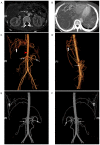
Duodenal angiomyolipoma with multiple systemic vascular malformations and aneurysms: A case report and literature review
- PMID: 29151912
- PMCID: PMC5680699
- DOI: 10.3892/ol.2017.7011
Duodenal angiomyolipoma with multiple systemic vascular malformations and aneurysms: A case report and literature review
Abstract
Angiomyolipomas (AMLs) are barely benign mesenchymal tumors that usually occur in the kidneys and may be associated with tuberous sclerosis complex (TSC). Extrarenal AMLs are markedly rare and infrequently observed in the duodenum. In the present case report, a 22-year-old female patient with duodenal AMLs presenting multiple systemic vascular malformations and aneurysms is described. The patient had a medical history of aneurysm rupture of the right subclavian artery and no other manifestation of TSC. Surgical intervention was performed. Following complete tumor resection, the patient declined to be treated further for vascular lesions. Pathological and immunohistochemical examination confirmed the diagnosis of duodenal AMLs. No tumor recurrence or progression of the vascular lesions was observed within 24 months of follow-up. This case report demonstrates the scarcity of duodenal AMLs with multiple systemic vascular malformations and aneurysms, which may be associated with novel gene mutations or TSC; however, further verification by gene sequencing is required.
Keywords: aneurysm; angiomyolipoma; duodenum; tuberous sclerosis; vascular malformation.
Figures


Similar articles
-
Renal manifestations of tuberous sclerosis complex: patients' and parents' knowledge and routines for renal follow-up - a questionnaire study.BMC Nephrol. 2018 Feb 13;19(1):39. doi: 10.1186/s12882-018-0835-3. BMC Nephrol. 2018. PMID: 29439672 Free PMC article.
-
The analysis of mutations and exon deletions at TSC2 gene in angiomyolipomas associated with tuberous sclerosis complex.Exp Mol Pathol. 2014 Dec;97(3):440-4. doi: 10.1016/j.yexmp.2014.09.013. Epub 2014 Oct 1. Exp Mol Pathol. 2014. PMID: 25281918
-
Sporadic versus Tuberous Sclerosis Complex-Associated Angiomyolipomas: Predictors for Long-Term Outcomes following Transcatheter Embolization.J Vasc Interv Radiol. 2016 Oct;27(10):1542-9. doi: 10.1016/j.jvir.2016.05.029. Epub 2016 Aug 10. J Vasc Interv Radiol. 2016. PMID: 27522275
-
Evolving Strategies in the Treatment of Tuberous Sclerosis Complex-associated Angiomyolipomas (TSC-AML).Urology. 2016 Mar;89:19-26. doi: 10.1016/j.urology.2015.12.009. Epub 2015 Dec 23. Urology. 2016. PMID: 26723178 Review.
-
Evidence-based protocol-led management of renal angiomyolipoma: A review of literature.Turk J Urol. 2021 Feb;47(Supp. 1):S9-S18. doi: 10.5152/tud.2020.20343. Epub 2020 Sep 21. Turk J Urol. 2021. PMID: 32966208 Free PMC article. Review.
Cited by
-
Tuberous sclerosis complex: a complex case.Cold Spring Harb Mol Case Stud. 2022 Apr 28;8(3):a006182. doi: 10.1101/mcs.a006182. Print 2022 Apr. Cold Spring Harb Mol Case Stud. 2022. PMID: 35483879 Free PMC article.
-
Brachial and subclavian arteries aneurysms due to tuberous sclerosis complex mechanisms - case report and literature review.Rom J Morphol Embryol. 2022 Jan-Mar;63(1):181-189. doi: 10.47162/RJME.63.1.19. Rom J Morphol Embryol. 2022. PMID: 36074682 Free PMC article. Review.
References
LinkOut - more resources
Full Text Sources
Other Literature Sources
