

Phloretin induces apoptosis of human esophageal cancer via a mitochondria-dependent pathway
- PMID: 29151915
- PMCID: PMC5680639
- DOI: 10.3892/ol.2017.7037
Phloretin induces apoptosis of human esophageal cancer via a mitochondria-dependent pathway
Abstract
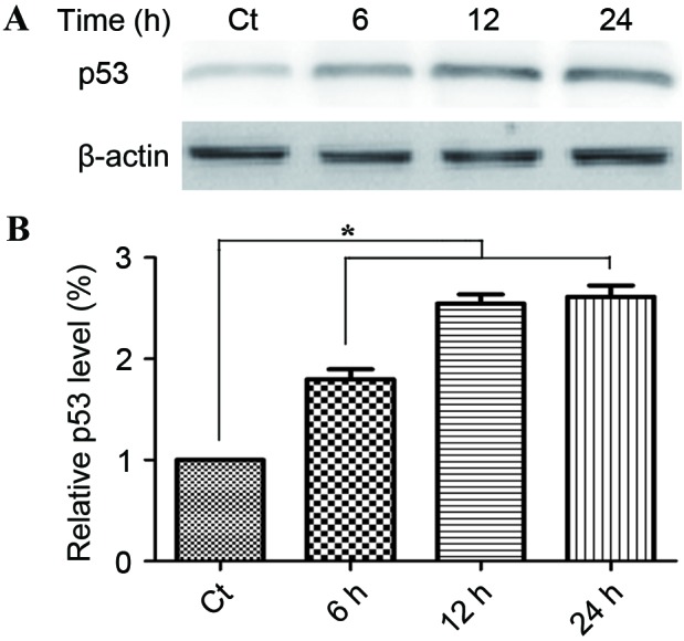
2,4,6-trihydroxy-3-(4-hydroxyphenyl)-propiophenone (phloretin) is found in apple tree leaves and the Manchurian apricot, and is a potent compound that exhibits anti-inflammatory, antioxidant and antitumor activities. However, the effect of phloretin on esophageal cancer cells is not well-defined. The present study aimed to examine whether and how phloretin induced apoptosis in human esophageal cancer cells. EC-109 cells were cultured in Dulbecco's modified Eagle's medium and incubated with 60, 70, 80, 90 and 100 µg/ml phloretin for 6, 12, 24 and 48 h. Cell proliferation was measured by an MTT assay. Cell apoptosis rate was measured using flow cytometric analysis subsequent to propidium iodide (PI) staining. The protein expression levels were determined by western blot analysis. It was found that phloretin significantly decreased viable cell numbers in a dose- and time-dependent manner and induced apoptosis in EC-109 cells. Additionally, phloretin exhibited potent anticancer activity in vitro, as evidenced by the downregulation of the anti-apoptosis-associated molecule B-cell lymphoma 2 (bcl-2) and an increase in the levels of the apoptosis-associated molecules bcl-2-like protein 4 and tumor protein p53. Phloretin treatment also affected the expression of apoptotic protease activating factor-1, the protein product of the direct binding of the inhibitor of apoptosis protein with low PI to the X-linked inhibitor of apoptosis protein. The present results indicated that phloretin may inhibit EC-109 cell growth by inducing apoptosis, which may be mediated through a mitochondria-dependent pathway.
Keywords: apoptosis; mitochondria-dependent pathway; phloretin.
Figures


Similar articles
-
Biochemical Basis of Anti-Cancer-Effects of Phloretin-A Natural Dihydrochalcone.Molecules. 2019 Jan 13;24(2):278. doi: 10.3390/molecules24020278. Molecules. 2019. PMID: 30642127 Free PMC article. Review.
-
Phloretin induces apoptosis in H-Ras MCF10A human breast tumor cells through the activation of p53 via JNK and p38 mitogen-activated protein kinase signaling.Ann N Y Acad Sci. 2009 Aug;1171:479-83. doi: 10.1111/j.1749-6632.2009.04692.x. Ann N Y Acad Sci. 2009. PMID: 19723092
-
Induction of apoptosis in HT-29 colon cancer cells by phloretin.J Med Food. 2007 Dec;10(4):581-6. doi: 10.1089/jmf.2007.116. J Med Food. 2007. PMID: 18158826
-
Cycloartobiloxanthone Induces Human Lung Cancer Cell Apoptosis via Mitochondria-dependent Apoptotic Pathway.In Vivo. 2018 Jan-Feb;32(1):71-78. doi: 10.21873/invivo.11206. In Vivo. 2018. PMID: 29275301 Free PMC article.
-
Phloretin, as a Potent Anticancer Compound: From Chemistry to Cellular Interactions.Molecules. 2022 Dec 12;27(24):8819. doi: 10.3390/molecules27248819. Molecules. 2022. PMID: 36557950 Free PMC article. Review.
Cited by
-
Amicis Omnia Sunt Communia: NF-κB Inhibition as an Alternative to Overcome Osteosarcoma Heterogeneity.Pharmaceuticals (Basel). 2024 Jun 5;17(6):734. doi: 10.3390/ph17060734. Pharmaceuticals (Basel). 2024. PMID: 38931401 Free PMC article. Review.
-
Stable Isotope Tracing Metabolomics to Investigate the Metabolic Activity of Bioactive Compounds for Cancer Prevention and Treatment.Cancers (Basel). 2020 Aug 3;12(8):2147. doi: 10.3390/cancers12082147. Cancers (Basel). 2020. PMID: 32756373 Free PMC article. Review.
-
Dihydrochalcones: Methods of Acquisition and Pharmacological Properties-A First Systematic Review.Molecules. 2019 Dec 5;24(24):4468. doi: 10.3390/molecules24244468. Molecules. 2019. PMID: 31817526 Free PMC article.
-
Onco-Preventive and Chemo-Protective Effects of Apple Bioactive Compounds.Nutrients. 2021 Nov 11;13(11):4025. doi: 10.3390/nu13114025. Nutrients. 2021. PMID: 34836282 Free PMC article. Review.
-
Unlocking the Therapeutic Potential of Natural Polyphenols in Esophageal Cancer.Curr Treat Options Oncol. 2025 Apr;26(4):278-290. doi: 10.1007/s11864-025-01308-6. Epub 2025 Mar 22. Curr Treat Options Oncol. 2025. PMID: 40120005 Review.
References
-
- Raymond DP, Seder CW, Wright CD, Magee MJ, Kosinski AS, Cassivi SD, Grogan EL, Blackmon SH, Allen MS, Park BJ, et al. Predictors of major morbidity or mortality after resection for esophageal cancer: A society of thoracic surgeons general thoracic surgery database risk adjustment model. Ann Thorac Surg. 2016;102:207–214. doi: 10.1016/j.athoracsur.2016.04.055. - DOI - PMC - PubMed
-
- Wu XD, Qin HY, Zhang JE, Zheng MC, Xin MZ, Liu L, Wu XJ, Jiang CN, Zhang MF. The prevalence and correlates of symptom distress and quality of life in Chinese oesophageal cancer patients undergoing chemotherapy after radical oesophagectomy. Eur J Oncol Nurs. 2015;19:502–508. doi: 10.1016/j.ejon.2015.02.010. - DOI - PubMed
LinkOut - more resources
Full Text Sources
Other Literature Sources
Research Materials
Miscellaneous