

Clinical Impact of microRNAs Associated With Cancer Stem Cells as a Prognostic Factor in Ovarian Carcinoma
- PMID: 29151939
- PMCID: PMC5687169
- DOI: 10.7150/jca.20348
Clinical Impact of microRNAs Associated With Cancer Stem Cells as a Prognostic Factor in Ovarian Carcinoma
Abstract
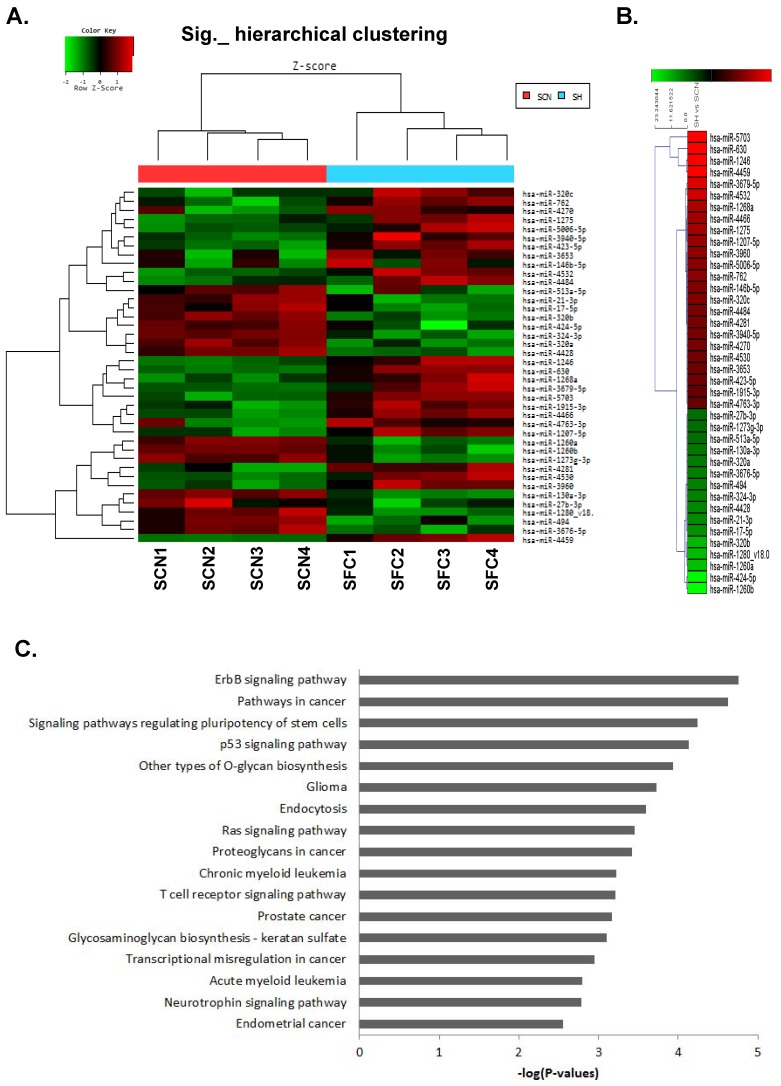
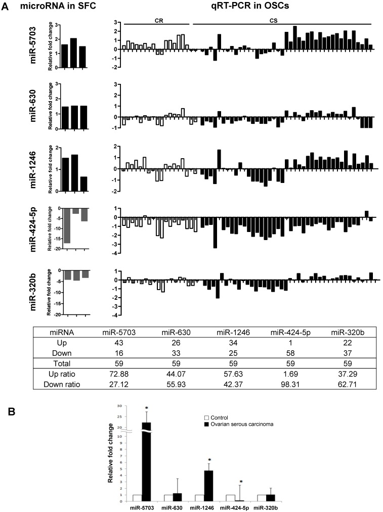
Background: Ovarian carcinoma is a highly lethal gynecological malignancy due to its frequent relapses and adoption of chemoresistance. To develop new biomarkers for disease progression in ovarian carcinoma, CSCs, which are considered to contribute to disease relapse and metastasis, were isolated from human ovarian carcinoma tissues, and differentially expressed microRNAs (miRNAs) in CSCs were identified and assessed the clinical implication of expression of these miRNAs. Methods: Primary cancer cells derived from human ovarian carcinomas were cultured and spheroid-forming cells (SFCs) were isolated. Profiles of miRNA expression in CSC-like SFCs were identified by miRNA microarray and the results were validated by quantitative real-time RT-PCR (qRT-PCR). We also assessed the correlations between miRNA expression levels and clinicopathological parameters in ovarian carcinomas. Results: Five miRNAs (miR-5703, miR-630, miR-1246, miR-424-5p, and miR-320b) were significantly dysregulated in CSC-like SFCs compared with primary cancer cells. The qRT-PCR showed that miR-5703 and miR-1246 expression was significantly higher in ovarian cancer cells than in normal control cells, whereas the miR-424-5p level was significantly lower. Decreased expression of miR-424-5p was significantly associated with distant metastasis in high stage (stage IIII & IV) carcinomas (35.5% vs. 72.2%, respectively, p=0.013) Conclusion: Taken together, miR-5703, miR-630, miR-1246, miR-424-5p, and miR-320b are useful markers for enriching ovarian CSCs. Decreased expression of miR-424-5p in ovarian carcinoma might be a putative biomarker for distant metastasis in ovarian carcinoma.
Keywords: Cancer stem cell; Ovarian carcinoma; miR-424-5p, Prognosis.; microRNA.
Conflict of interest statement
Competing Interests: The authors have declared that no competing interest exists.
Figures
References
-
- Jemal A, Thomas A, Murray T, Thun M. Cancer statistics, 2002. CA Cancer J Clin. 2002;52(1):23–47. - PubMed
-
- McGuire WP, Hoskins WJ, Brady MF, Kucera PR, Partridge EE, Look KY, Clarke-Pearson DL, Davidson M. Cyclophosphamide and cisplatin versus paclitaxel and cisplatin: a phase III randomized trial in patients with suboptimal stage III/IV ovarian cancer (from the Gynecologic Oncology Group) Semin Oncol. 1996;23(5 Suppl 12):40–47. - PubMed
-
- Lapidot T, Sirard C, Vormoor J, Murdoch B, Hoang T, Caceres-Cortes J, Minden M, Paterson B, Caligiuri MA, Dick JE. A cell initiating human acute myeloid leukaemia after transplantation into SCID mice. Nature. 1994;367(6464):645–648. - PubMed
-
- Kim CF, Dirks PB. Cancer and stem cell biology: how tightly intertwined? Cell Stem Cell. 2008;3(2):147–150. - PubMed
-
- Ahmed N, Abubaker K, Findlay J, Quinn M. Cancerous ovarian stem cells: obscure targets for therapy but relevant to chemoresistance. J Cell Biochem. 2013;114(1):21–34. - PubMed
LinkOut - more resources
Full Text Sources
Other Literature Sources
