. 2017 Sep 15;8(50):87582-87597.
doi: 10.18632/oncotarget.20908. eCollection 2017 Oct 20.

Myxinidin2 and myxinidin3 suppress inflammatory responses through STAT3 and MAPKs to promote wound healing

Affiliations

Myxinidin2 and myxinidin3 suppress inflammatory responses through STAT3 and MAPKs to promote wound healing

Hyo Mi Han et al. Oncotarget. .

Abstract

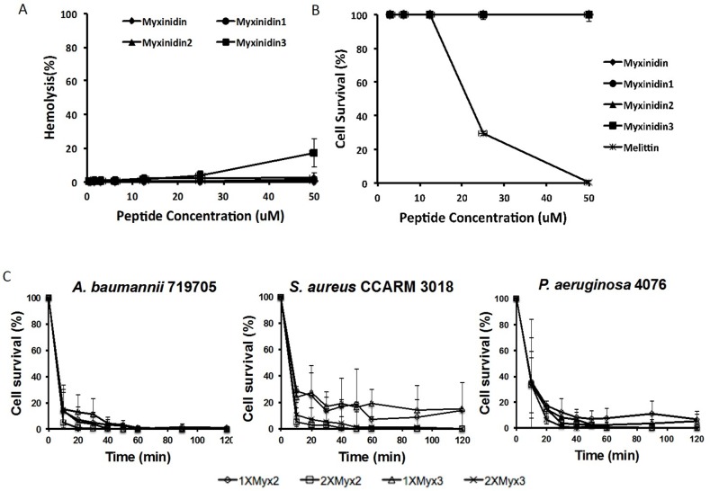
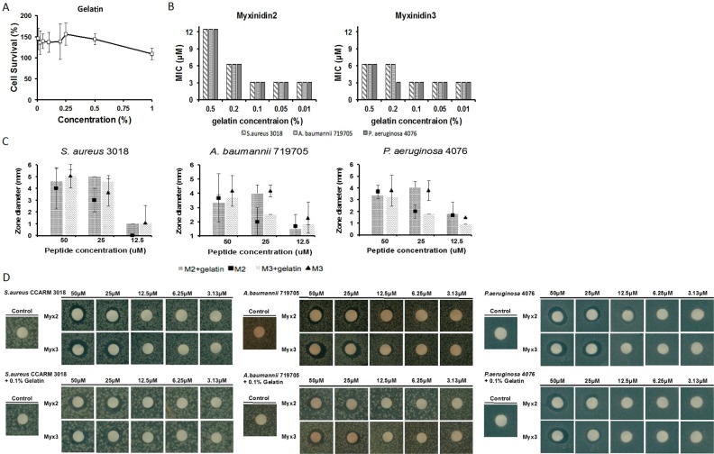
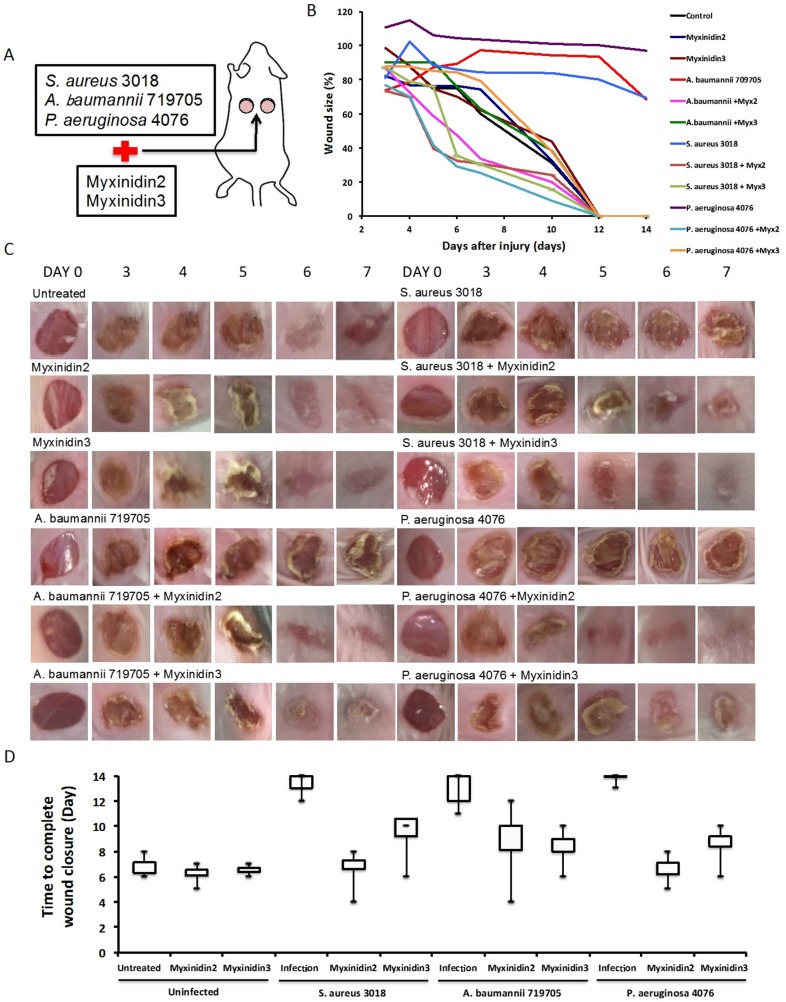
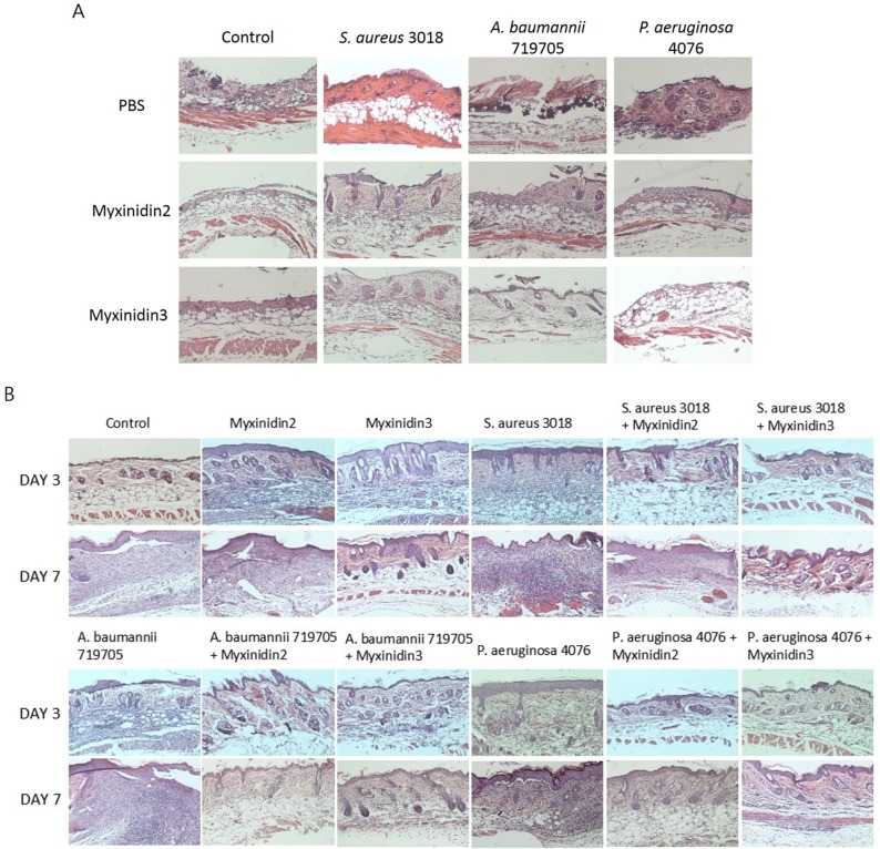
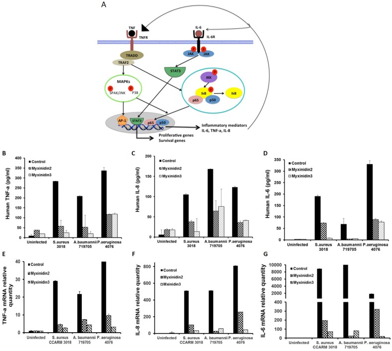
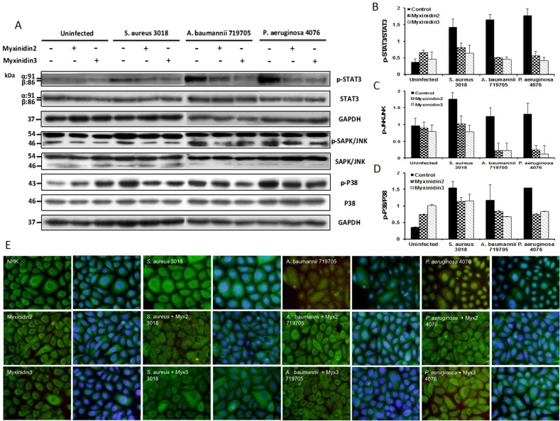
Skin wounds are continuously exposed to bacteria and can easily become infected. Infected wounds require antibiotic treatment, and infections caused by drug-resistant bacteria are an important public health problem. Antimicrobial peptides have broad-spectrum antibacterial activity, induce little or no drug resistance and may be suitable for treating skin infections caused by drug-resistant bacteria. We previously reported the design and function of myxinidin and myxinidin analogues. Here we showed that myxinidin2 and myxinidin3 exhibit antimicrobial and anti-biofilm activities against antibiotic-resistant Staphylococcus aureus, Acinetobacter baumannii, and Pseudomonas aeruginosa in high salt environments and in gelatin. Moreover, these peptides facilitated infected wound healing by decreasing inflammation through suppression of IL-6, IL-8, and TNF-α and regulation of downstream mediators such as STAT3, p38, JNK, and EGFR. In a mouse skin wound model infected with antibiotic-resistant bacteria, myxinidin2 and myxinidin3 eliminated the infection and enhanced wound healing. We therefore propose the use of these peptides for treating infected wounds and burns.

Keywords: MAPKs; STAT3; antimicrobial peptide; myxinidin; wound healing.


Conflict of interest statement

CONFLICTS OF INTEREST The authors declared no conflicts of interest.

Figures

Figure 1
Figure 1. Antibiotic killing kinetics of myxinidin2 and myxinidin3
(A) Hemolytic activity of the indicated peptides against RBCs. Release of hemoglobin was measured at 414 nm. (B) Cytotoxicity activity of the peptides assessed using by MTT assays. Normal human keratinocytes were used to evaluate cytotoxicity. Measured was the absorbance at 570 and 650 nm. (C) Time-to-kill kinetics curve for the indicated peptides against S. aureus 3018, A. baumannii 719705 and P. aeruginosa 4076. Each experiment was performed in triplicate, and the value shown is the average of each experiment.
Figure 2
Figure 2. Anti-biofilm activity of myxinidin2 and myxinidin3
Inhibition of biofilm formation was examined at the indicated concentrations of the peptides (A) 6.25 μM, (B) 12.5 μM, (C) 25 μM, (D) 50 μM. Each experiment was performed in triplicate, with at least three independent experiments.
Figure 3
Figure 3. Antimicrobial activity of myxinidin2 and myxinidin3 in gelatin
(A) Cytotoxicity of 0% to 1% gelatin against normal human keratinocytes. (B) Sustained antimicrobial effect at each gelatin concentration was verified by determining the MIC. (C) and (D) Disc assays showing the antibacterial effects of myxinidin2 and myxinidin3 in 0.1% gelatin. Each experiment was performed in triplicate, with at least three independent experiments.
Figure 4
Figure 4. Effects of myxinidin2 and myxinidin3 on migration of infected keratinocytes
(A) Graph showing the effects of the indicated peptides on wound area coverage by infected keratinocytes at the indicated times. (B) Visualization of wound closing through keratinocyte migration at the indicated times after wounding. Dotted lines denote the wound. Cells were infected with the indicated bacterial with or without myxinidin2 and myxinidin3 treatment. (C) and (D) Western blot analysis showing that myxinidin2 and myxinidin3 stimulated EGFR phosphorylation. (E) Immunofluorescent staining revealed activation of EGFR by myxinidin2 and myxinidin3 at the surface of infected keratinocytes. EGFR was detected using anti-EGFR antibody (green). The nuclei were stained with Hoechst (blue). Each experiment was performed in triplicate, with at least three independent experiments.
Figure 5
Figure 5. Effect of myxinidin2 and myxinidin3 on wound healing in vivo
(A) Schematic illustration of wound healing model. (B) Percent change in wound size over time. (C) Images of wounds 0, 3, 4, 5, 6, 7 days after injury. (D) Time to complete wound closure in each group. Values shown are the means of three individual experiments.
Figure 6
Figure 6. Histological evaluation of infected wound sections
(A) H&E stained sections of infected wounds, with and without myxinidin2/3 treatment. (B) H&E stained section of infected wounds 3 and 7 days after injury, with and without myxinidin2/3 treatment.
Figure 7
Figure 7. Model of inflammatory signal pathways in keratinocytes
(A) Normal human keratinocytes were infected with S. aureus CCARM 3018, A. baumannii 79705 or P. aeruginosa 4076 and then treated with myxinidin2/3. Expression levels of cytokines and chemokines were quantified using ELISAs. (B) TNF-α, (C) IL-8, (D) IL-6. Levels of cytokine and chemokine mRNAs were quantified using qRT-PCR. (E) TNF-α, (F) IL-8, (G) IL-6. Each experiment was performed in triplicate, with at least three independent experiments.
Figure 8
Figure 8
(A) Western blot analysis of the effects of myxinidin2 and myxinidin3 on levels of phosphorylated STAT3 and MAPK (SAPK/JNK and P38). Cells were pre-infected with the indicated bacteria before addition of the peptides. The results are configured as bar graphs in (B), (C) and (D). (E) Myxinidin2 and myxinidin3 inhibit bacteria-induced NF-kB translocation in normal human keratinocytes. NF-kB was detected using a polyclonal anti-NF-kB p65 antibody. Its intracellular localization (green) was compared with Hoechst stained nuclei (blue). Each experiment was performed in triplicate, with at least three independent experiments.

Similar articles

Cited by

References

    1. Kumar A, Zhang J, Yu FS. Innate immune response of corneal epithelial cells to Staphylococcus aureus infection: role of peptidoglycan in stimulating proinflammatory cytokine secretion. Invest Ophthalmol Vis Sci. 2004;45:3513–22. - PMC - PubMed
    1. March C, Regueiro V, Llobet E, Moranta D, Morey P, Garmendia J, Bengoechea JA. Dissection of host cell signal transduction during Acinetobacter baumannii-triggered inflammatory response. PLoS One. 2010;5:e10033. - PMC - PubMed
    1. Liu Y, Zhou Q, Wang Y, Liu Z, Dong M, Wang Y, Li X, Hu D. Negative pressure wound therapy decreases mortality in a murine model of burn-wound sepsis involving Pseudomonas aeruginosa infection. PLoS One. 2014;9:e90494. - PMC - PubMed
    1. Hoque J, Adhikary U, Yadav V, Samaddar S, Konai MM, Prakash RG, Paramanandham K, Shome BR, Sanyal K, Haldar J. Chitosan derivatives active against multidrug-resistant bacteria and pathogenic fungi: in vivo evaluation as topical antimicrobials. Mol Pharm. 2016;13:3578–89. - PubMed
    1. Dalac S, Sigal L, Addala A, Chahim M, Faivre-Carrere C, Lemdjadi Z, Bohbot S. Clinical evaluation of a dressing with poly absorbent fibres and a silver matrix for managing chronic wounds at risk of infection: a non comparative trial. J Wound Care. 2016;25:531–8. - PubMed