Editorial
. 2017 Oct;6(5):312-316.
doi: 10.21037/hbsn.2016.12.04.

Robotic transduodenal excision of ampullary tumour

Affiliations
Editorial

Robotic transduodenal excision of ampullary tumour

Francis C H Wong et al. Hepatobiliary Surg Nutr. 2017 Oct.

Abstract

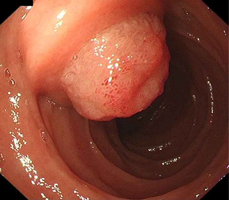
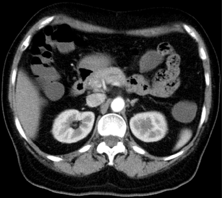
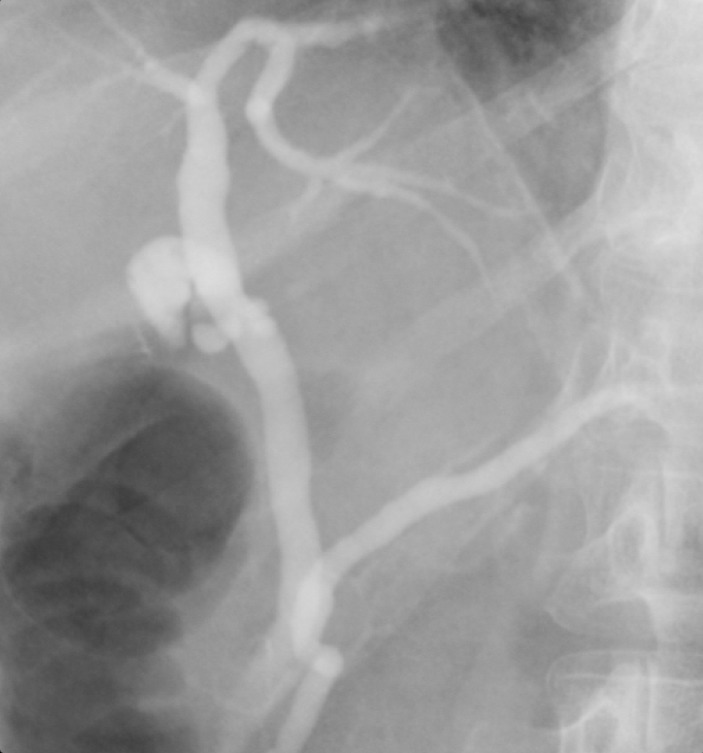
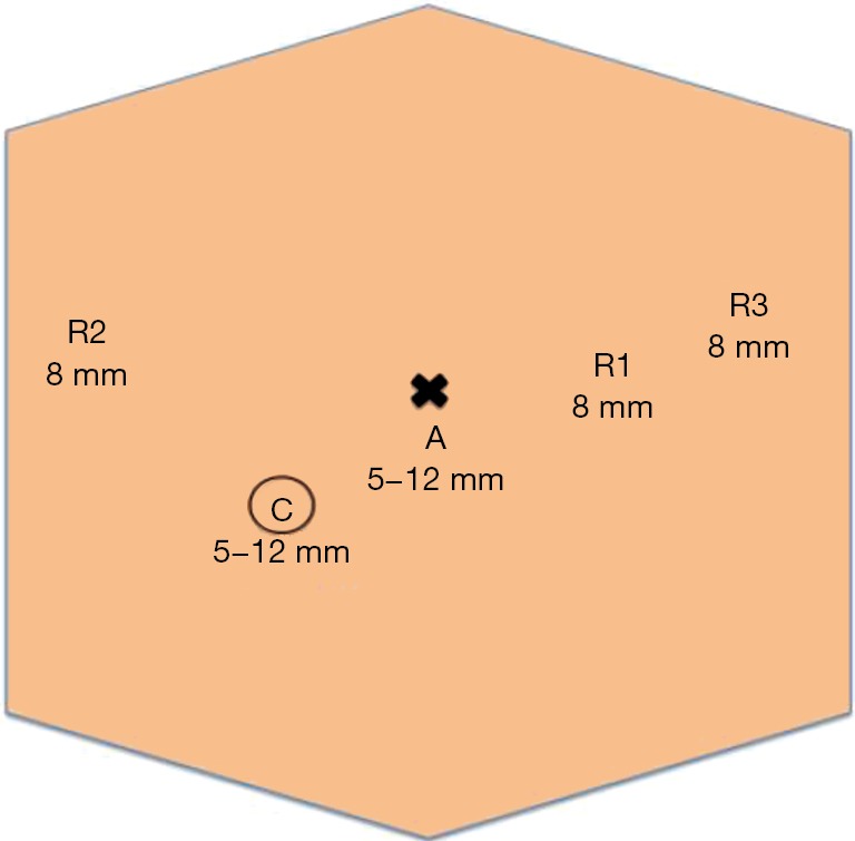
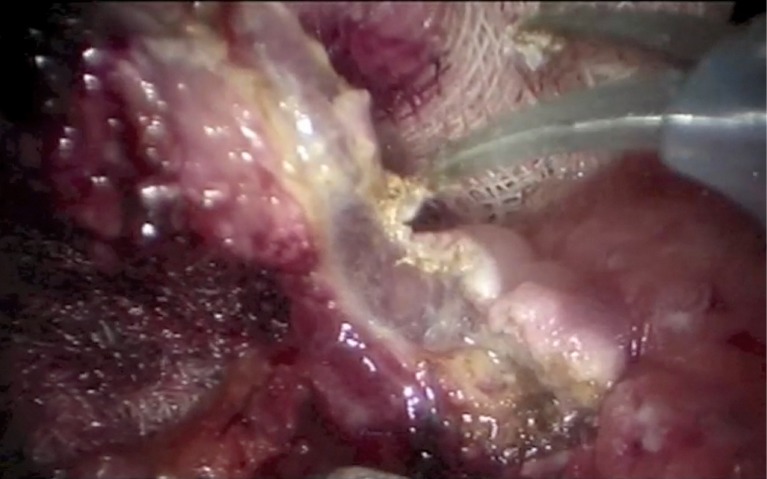
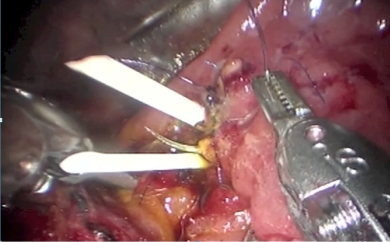
Ampullary tumours are uncommon lesions with potential risk of malignancy. The management is excision by either endoscopic ampullectomy, pancreaticoduodenectomy or transduodenal excision. Endoscopic ampullectomy offers a less invasive approach, whereas pancreaticoduodenectomy allows radical excision of the tumour. They both carry their own limitations. Transduodenal excision of ampullary tumour offers significantly lower risks with low recurrence rate, and can be offered for benign or early grade tumours. Limited cases of laparoscopic transduodenal excision of ampullary tumours were reported in the literature, probably due to the technical difficulties in performing the operation, especially during the reconstruction of the pancreaticobiliary ducts with the laparoscopic approach. In the era with robotic surgical systems, the technical demanding procedures are greatly facilitated. Hence, we are writing to report a case of transduodenal excision of ampullary tumour with the robot assisted laparoscopic approach.

Keywords: Robotic; ampulla; operative technique; transduodenal excision; tumour.


Conflict of interest statement

Conflicts of Interest: The authors have no conflicts of interest to declare.

Figures

Figure 1
Figure 1
Oesophagogastroduodenoscopy (OGD) showing a villous growth over ampulla of Vater.
Figure 2
Figure 2
Contrast enhanced CT scan showing no enhancing lesion. CT, computed tomography.
Figure 3
Figure 3
PET scan showing no uptake over the papillary region. PET, positron emission tomography.
Figure 4
Figure 4
ERCP in 1 year post-operatively showing no biliary or pancreatic ductal strictures. ERCP, endoscopic retrograde cholangiopancreatography.
Figure 5
Figure 5
Robotic arms, camera and assistant port placements. C, camera port; R, robotic arm; A, assistant port.
Figure 6
Figure 6
Excision of the ampullary tumour after raised by submucosal adrenaline saline injection.
Figure 7
Figure 7
Insertion of stent into pancreatic and bile duct individually and anchored with 3/0 Monocryl sutures.

References

    1. Albores-Saavedra J, Schwartz AM, Batich K, et al. Cancers of the ampulla of vater: demographics, morphology, and survival based on 5,625 cases from the SEER program. J Surg Oncol 2009;100:598-605. 10.1002/jso.21374 - DOI - PubMed
    1. Schneider L, Contin P, Fritz S, et al. Surgical ampullectomy: an underestimated operation in the era of endoscopy. HPB (Oxford) 2016;18:65-71. 10.1016/j.hpb.2015.07.004 - DOI - PMC - PubMed
    1. Beger HG, Treitschke F, Gansauge F, et al. Tumor of the ampulla of Vater: experience with local or radical resection in 171 consecutively treated patients. Arch Surg 1999;134:526-32. 10.1001/archsurg.134.5.526 - DOI - PubMed
    1. Rosen M, Zuccaro G, Brody F. Laparoscopic resection of a periampullary villous adenoma. Surg Endosc 2003;17:1322-3. 10.1007/s00464-002-4527-4 - DOI - PubMed
    1. Zhang RC, Xu XW, Wu D, et al. Laparoscopic transduodenal local resection of periampullary neuroendocrine tumor: a case report. World J Gastroenterol 2013;19:6693-8. 10.3748/wjg.v19.i39.6693 - DOI - PMC - PubMed

Publication types

LinkOut - more resources