. 2017 Dec;16(6):9457-9463.
doi: 10.3892/mmr.2017.7823. Epub 2017 Oct 19.

Naringin protects myocardial cells from doxorubicin‑induced apoptosis partially by inhibiting the p38MAPK pathway

Affiliations

Naringin protects myocardial cells from doxorubicin‑induced apoptosis partially by inhibiting the p38MAPK pathway

Chun-Yan Jian et al. Mol Med Rep. 2017 Dec.

Abstract

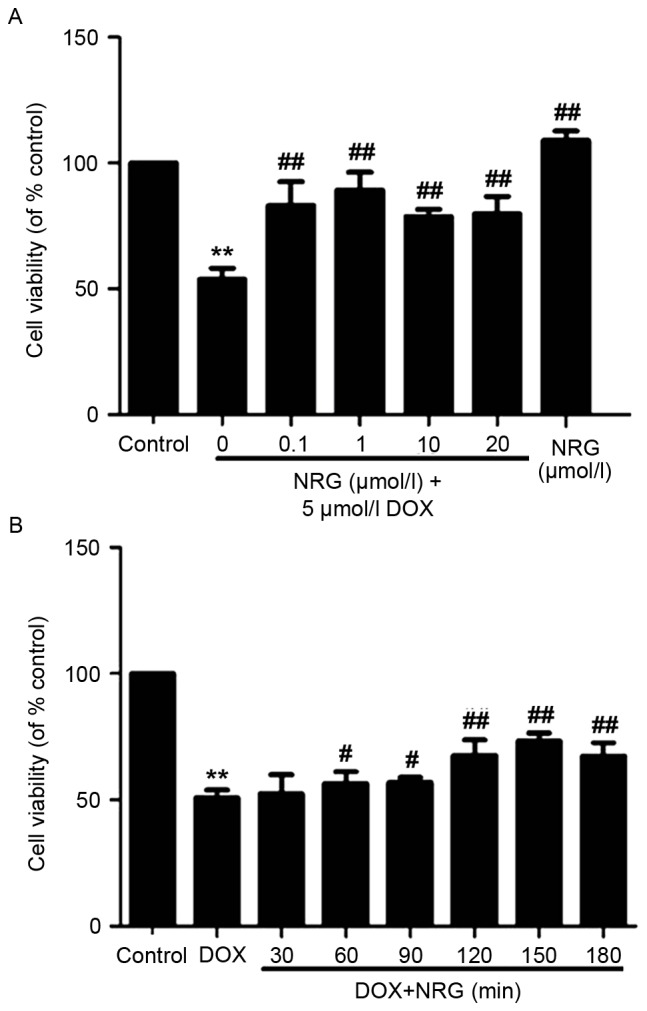
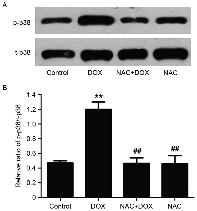
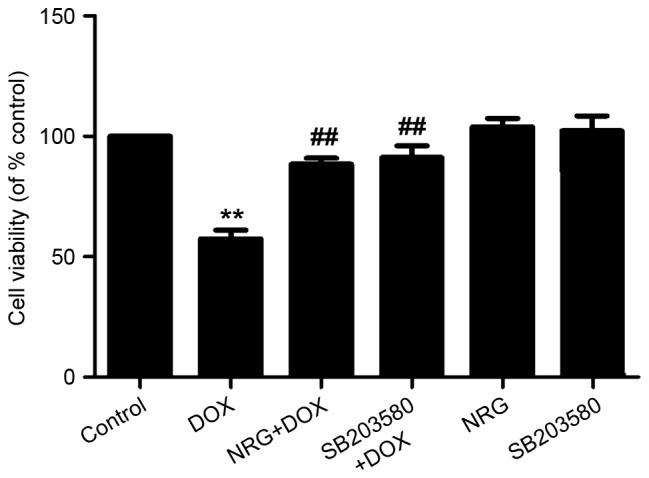
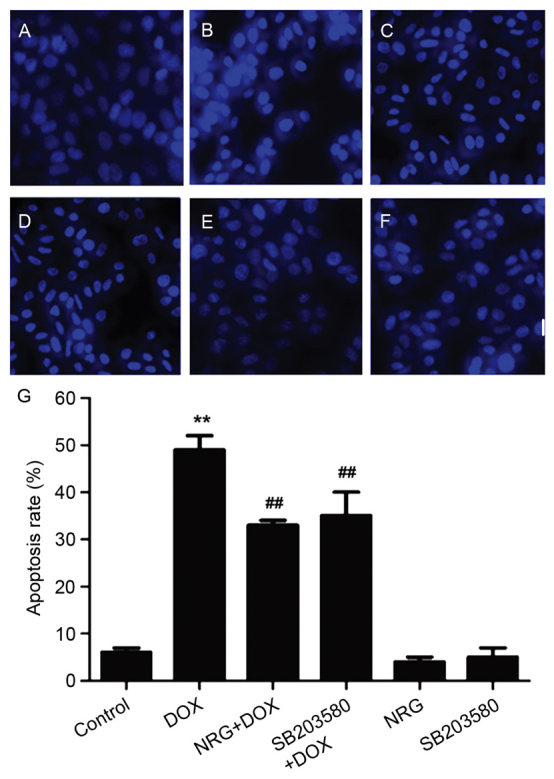
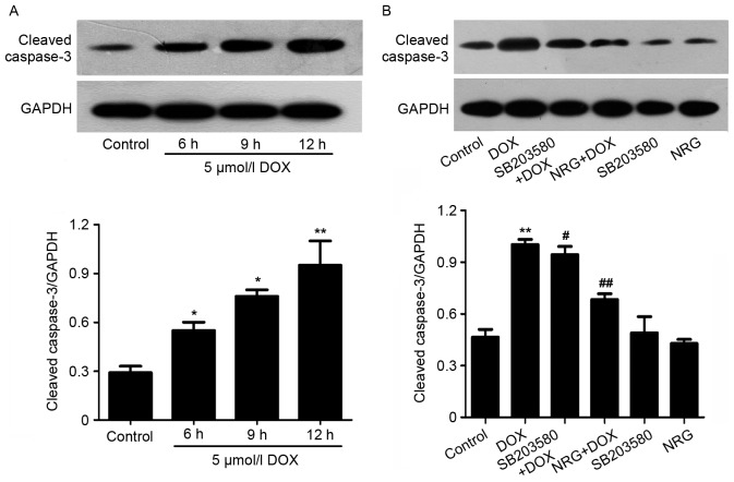
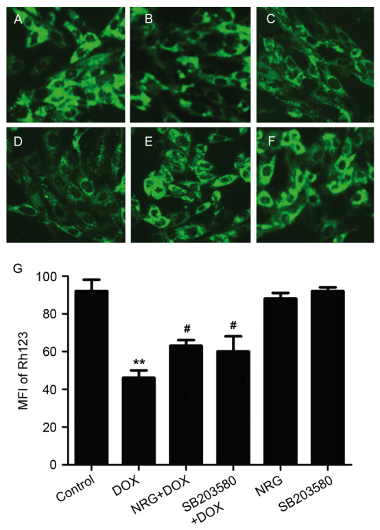
Doxorubicin (DOX) has been widely used to treat cancers as a first‑line antitumor drug. However, it causes severe, irreversible, dose‑dependent cardiotoxicity. To evaluate the protective effects of naringin (NRG) on cardiotoxicity, the authors investigated the molecular mechanism of the p38MAPK signaling pathway. H9c2 cells were treated for 24 h by using 5 µmol/l DOX without or with being pretreated by 1 µM NRG for 150 min or by 3 µM SB203580 for 60 min. Cell viability was detected by cell counting kit‑8 assay. Intracellular reactive oxygen species (ROS) levels were detected based on the oxidative conversion of 2',7'‑dichlorfluorescein‑diacetate (cell‑permeable) to dichlorofluorescein (fluorescent). The expression of p38MAPK was determined by western blotting. The expression level of p‑p38MAPK in H9c2 cells, which was significantly increased by exposure to 5 µM DOX for 60 min (P<0.01), was significantly decreased by pretreatment with 1 µM NRG for 150 min beforehand (P<0.01). The viability of H9c2 cells pretreated for 150 min with 1 µM NRG was significantly enhanced compared with that using DOX directly (P<0.01). Intracellular ROS levels were significantly reduced by being pretreated with 1 µM NRG for 150 min or with 3 µM SB203580 for 60 min before the cells were exposed to 5 µM DOX. Collectively, NRG protected H9c2 cells against the cardiotoxicity induced by DOX through suppressing the expression and activity of the p38MAPK pathway. The findings provided valuable evidence for the possible use of NRG to relieve DOX‑induced cardiotoxicity.


Figures

Figure 1.
Figure 1.
Influence of NRG on the viability of H9c2 cells. (A) Cells were pretreated with NRG at indicated concentrations for 120 min prior to exposure to 5 µmol/l DOX for 24 h. (B) Cells were pretreated with 1 µmol/l NRG for indicated times prior to DOX exposure. CCK-8 assay was conducted to measure cell viability. The data are expressed as mean ± standard error of the mean (n=3). **P<0.01 vs. Control; #P<0.05, ##P<0.01 vs. DOX-treated group alone. NRG, naringin; DOX, doxorubicin.
Figure 2.
Figure 2.
Influence of NRG on p-p38MAPK expression. (A) H9c2 cells were treated for 60 min with 5 µM DOX in the presence or absence of 150 min of pretreatment by using 1 µM NRG, and p38MAPK expression was detected by western blot analysis. (B) The data are expressed as mean ± standard error of the mean (n=3). **P<0.01 vs. Control; ##P<0.01 vs. DOX-treated group. NRG, naringin; DOX, doxorubicin; MAPK, mitogen-associated protein kinase.
Figure 3.
Figure 3.
Roles of oxidative stress in DOX-induced upregulation of p-p38MAPK. (A) H9c2 cells were treated for 60 min with 5 µM DOX in the presence or absence of 60 min of pretreatment by using 1,000 µM NAC, and p38MAPK expression was detected by western blot analysis. (B) The data are expressed as mean ± standard error of the mean (n=3). **P<0.01 vs. Control; ##P<0.01 vs. DOX-treated group. NAC, N-acetyl-L-cysteine; DOX, doxorubicin.
Figure 4.
Figure 4.
Effects of NRG and p38MAPK inhibitor on cytotoxicity induced by DOX. H9c2 cells were treated for 24 h with 5 µmol/l DOX in the presence or absence of 150 min of pretreatment by using 1 µM NRG for or 60 min of pretreatment by using 3 µM SB203580. The viability was then detected with Cell Counting kit-8 assay. The data are expressed as mean ± standard error of the mean (n=3). **P<0.01 vs. Control, ##P<0.01 vs. DOX-treated group. NRG, naringin; DOX, doxorubicin.
Figure 5.
Figure 5.
Effects of NRG and p38MAPK inhibitor on DOX-induced apoptosis of H9c2 cells. Following different treatments, H9c2 cells were stained by Hoechst 33258, the apoptosis of which was observed with fluorescence imaging. (A) Control group. (B) The cells were exposed for 24 h to DOX at 5 µM. (C) The cells were pretreated for 150 min by using 1 µM NRG prior to 24 h of exposure to DOX at 5 µM. (D) The cells were treated for 60 min by using 3 µM SB203580 prior to 24 h of exposure to DOX at 5 µM. (E) The cells were treated for 150 min by using 1 µM NRG and cultured for 24 h. (F) The cells were treated for 60 min with 3 µM SB203580 and cultured for 24 h. (G) Apoptotic rate was analyzed by the cell counter of ImageJ software. The data are expressed as mean ± standard error of the mean (n=3). Magnification, ×400. **P<0.01 vs. Control; ##P<0.01 vs. DOX-treated group. NRG, naringin; DOX, doxorubicin.
Figure 6.
Figure 6.
Western blot analyses. (A) Effects of DOX on the expression of cleaved caspase-3 after 6–12 h of H9c2 cell exposure to DOX at 5 µM. (B) Effects of NRG and p38MAPK inhibitor on DOX-induced increase in cleaved caspase-3 expression. The cells were treated for 12 h by using 5 µmol/l DOX in the presence or absence of 150 min of pretreatment by 1 µM NRG or 60 min of pretreatment by 3 µM SB203580. The data were quantified by ImageJ software using densitometric analysis and expressed as mean ± standard error of the mean (n=3). *P<0.05, **P<0.01 vs. Control; ##P<0.01, #P<0.05 vs. DOX-treated group, NRG, naringin; DOX, doxorubicin.
Figure 7.
Figure 7.
Immunofluorescence staining of reactive oxygen species in differently treated H9c2 cells. (A) Control group. (B) The cells were exposed for 24 h to DOX at 5 µM. (C) The cells were pretreated for 150 min by NAG at 1 µM before 24 h of exposure to DOX at 5 µM. (D) The cells were pretreated for 60 min by 3 µM SB203580 before 24 h of exposure to DOX at 5 µM. (E) The cells were treated for 150 min by 1 µM NRG and cultured for 24 h. (F) The cells were treated for 60 min by 3 µM SB203580 and cultured for 24 h. (G) Quantified MFI of DCF in A-F by Image J software. The data are expressed as mean ± standard error of the mean (n=3). Magnification, ×400. *P<0.05 vs. Control; #P<0.05 vs. DOX-treated group. DOX, doxorubicin; NAC, N-acetyl-L-cysteine; NRG, naringin; DCF, dichlorofluorescein; MFI, mean fluorescence intensity.
Figure 8.
Figure 8.
Immunofluorescence staining of mitochondrial membrane potential in differently treated H9c2 cells. (A) Control group. (B) The cells were treated for 24 h by DOX at 5 µM. (C) The cells were pretreated for 150 min by 1 µM NRG before 24 h of exposure to DOX at 5 µM. (D) The cells were pretreated for 60 min by 3 µM SB203580 before 24 h of exposure to DOX at 5 µM. (E) The cells were treated for 30 min by 1 µM NRG and cultured for 24 h. (F) The cells were treated for 60 min by 3 µM SB203580 and cultured for 24 h. (G) Quantified MFI of Rh123 in A-F by Image J software. The data are expressed as mean ± standard error of the mean (n=5). **P<0.01 vs. control; #P<0.05 vs. DOX-treated group. DOX, doxorubicin; NRG, naringin; MFI, mean fluorescence intensity; Rh123, rhodamine123.

References

    1. Bai J, Ma M, Cai M, Xu F, Chen J, Wang G, Shuai X, Tao K. Inhibition enhancer of zeste homologue 2 promotes senescence and apoptosis induced by doxorubicin in p53 mutant gastric cancer cells. Cell Prolif. 2014;47:211–218. doi: 10.1111/cpr.12103. - DOI - PMC - PubMed
    1. Hrdina R, Gersl V, Klimtová I, Simůnek T, Machácková J, Adamcová M. Anthracycline-induced cardiotoxicity. Acta Medica (Hradec Kralove) 2000;43:75–82. - PubMed
    1. Scully RE, Lipshultz SE. Anthracycline cardiotoxicity in long-term survivors of childhood cancer. Cardiovasc Toxicol. 2007;7:122–128. doi: 10.1007/s12012-007-0006-4. - DOI - PubMed
    1. Ludke AR, Al-Shudiefat AA, Dhingra S, Jassal DS, Singal PK. A concise description of cardioprotective strategies in doxorubicin-induced cardiotoxicity. Can J Physiol Pharmacol. 2009;87:756–763. - PubMed
    1. Alkreathy H, Damanhouri ZA, Ahmed N, Slevin M, Ali SS, Osman AM. Aged garlic extract protects against doxorubicin-induced cardiotoxicity in rats. Food Chem Toxicol. 2010;48:951–956. doi: 10.1016/j.fct.2010.01.005. - DOI - PubMed

MeSH terms