Review
. 2018 Jan 18;25(1):36-49.
doi: 10.1016/j.chembiol.2017.10.007. Epub 2017 Nov 16.

APOBEC Enzymes as Targets for Virus and Cancer Therapy

Affiliations
Review

APOBEC Enzymes as Targets for Virus and Cancer Therapy

Margaret E Olson et al. Cell Chem Biol. .

Abstract

Human DNA cytosine-to-uracil deaminases catalyze mutations in both pathogen and cellular genomes. APOBEC3D, APOBEC3F, APOBEC3G, and APOBEC3H restrict human immunodeficiency virus 1 (HIV-1) infection in cells deficient in the viral infectivity factor (Vif), and have the potential to catalyze sublethal levels of mutation in viral genomes in Vif-proficient cells. At least two APOBEC3 enzymes, and in particular APOBEC3B, are sources of somatic mutagenesis in cancer cells that drive tumor evolution and may manifest clinically as recurrence, metastasis, and/or therapy resistance. Consequently, APOBEC3 enzymes are tantalizing targets for developing chemical probes and therapeutic molecules to harness mutational processes in human disease. This review highlights recent efforts to chemically manipulate APOBEC3 activities.

Keywords: APOBEC; cancer; chemical probes; cytosine-to-uracil; mutation; virus.


Figures

Figure 1
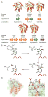
Figure 1. Structure, Organization, and Enzymatic Activity of Human A3 Enzymes
(A) The seven human A3 family members are distinguished by their number of structural domains (represented as one or two arrows), the phylogenetic grouping of each domain (Z1, green; Z2, orange; Z3, blue), and sub-cellular localization. Rendered X-ray structures (PyMOL) depict the catalytic domains of A3B (pdb 5CQD), A3F (pdb 5HX5) and A3G (pdb 3V4K), and the full-length structures of A3A (pdb 5SWW) and A3C (pdb 3VM8) (Li et al., 2012; Shaban et al., 2016; Shi et al., 2017; Shi et al., 2015). Structurally, each A3 domain has six alpha helices (red) and five beta strands (yellow). The flexible and variable loops are depicted in green. The active site coordinates a single zinc ion (grey sphere). (B) The proposed mechanism of A3-mediated ssDNA C-to-U deamination. (C) A3A-ssDNA X-ray co-crystal structure (pdb 5SWW) that shows the DNA substrate binds in a U-shaped confirmation with the −1 based flipped out of the active site and the target DNA cytosine interacting with the catalytic zinc (Shi et al., 2017). The −1 base forms specificity-conferring H-bonding contacts with residues of A3A.
Figure 2
Figure 2. Model for A3-Mediated HIV-1 Restriction
In an infected cell, a sublethal number of A3s incorporate into budding viral particles and hitch-hike to virus naïve cells. HIV-1 modulates A3 expression through Vif, which forms an E3 ubiquitin ligase complex that polyubiquitinates A3s and triggers their degradation at the 26S proteasome. The A3s restrict viral replication through both deamination-dependant mutagenesis and deamination-independent RT inhibition mechanisms. Adapted from (Harris and Dudley, 2015).
Figure 3
Figure 3. Therapeutic Strategies to Target the A3-Vif Interface
(A) In a clinical infection, A3-catalyzed restriction is inhibited by Vif-mediated ubiquitylation and proteasomal degradation. Despite this inhibition, a sublethal level of A3 deamination is observed. Thus, the activities of Vif and the A3s are balanced to achieve optimum viral fitness. (B) Therapy by hypermutation: This therapeutic strategy seeks to inhibit Vif and/or block the Vif-A3 interface thereby reinstating the restrictive capabilities of the A3s (Haché et al., 2006). Upon Vif inhibition, the relevant A3s can lethally mutate the viral genome. (C) Therapy by hypomutation: Inhibition of the A3s may deprive the virus of a needed mutation source. Inhibiting viral fitness may enable immunological or antiretroviral HIV-1 clearance (Harris, 2008).
Figure 4
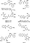
Figure 4
Chemical Structures of Small Molecules that Function through “Therapy by Hypermutation”.
Figure 5
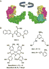
Figure 5. Candidate Inhibitors of Vif Interaction Surfaces
(A) X-ray structure of the Vif/CBF-β/CUL5/ELOB/ELOC complex (pdb 4N9F) (Guo et al., 2014). Color Scheme: Vif (magenta), CBF-β (yellow), CUL5 NTD (green), ELOC (orange), ELOB (cyan), Zn (grey). (B) Chemical structures of VEC-5, a putative Vif-ELOC PPI inhibitor, SN-1 and -2 and Baculiferin L and M, Vif-A3 PPI inhibitors.
Figure 6
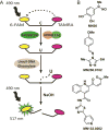
Figure 6. HTS for Small Molecules that Function through “Therapy by Hypermutation”
(A) Schematic of a fluorescence-based C-to-U deamination assay for high-throughput screening. Uninhibited A3-catalyzed deamination results in a high fluorescence readout, while potent A3 inhibition reads as background fluorescence. (B) Chemical structures of published A3G inhibitors.
Figure 7
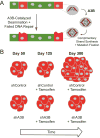
Figure 7. Model Depicting the Impact of A3B-Catalyzed Base Substitution Mutation in Cancer
(A) Mutation catalyzed by A3B can accelerate tumor cell growth, metastasis, and the development of therapeutic resistance. A3B preferentially deaminates DNA cytosines in a 5′-TCA context. The resulting uracil templates the insertion of adenine during complementary strand synthesis and uracil base excision repair will convert the U-A base pair to T-A (a C-to-T transition mutation). (B) Schematic representation of tumor volume during an A3B knockdown study in an ER+ breast cancer xenograft model (Law et al., 2016). At 50 days, mice were injected with tumor cells expressing a shRNA control or a shRNA to knockdown endogenous A3B. At 125 days, TAM treatment was initiated to suppress growth of similarly sized tumors. However, by 300 days, most of the control (A3B expressing) tumors had become resistant to TAM therapy, whereas the growth of most of the A3B depleted tumors was still suppressed. This study demonstrates that A3B contributes to the development of tamoxifen resistance.

References

    1. Albin JS, Harris RS. Interactions of host APOBEC3 restriction factors with HIV-1 in vivo: implications for therapeutics. Expert Rev Mol Med. 2010;12:e4. - PMC - PubMed
    1. Alce TM, Popik W. APOBEC3G is incorporated into virus-like particles by a direct interaction with HIV-1 Gag nucleocapsid protein. J Biol Chem. 2004;279:34083–34086. - PubMed
    1. Alexandrov LB, Nik-Zainal S, Wedge DC, Aparicio SA, Behjati S, Biankin AV, Bignell GR, Bolli N, Borg A, Borresen-Dale AL, et al. Signatures of mutational processes in human cancer. Nature. 2013;500:415–421. - PMC - PubMed
    1. Ali A, Wang J, Nathans RS, Cao H, Sharova N, Stevenson M, Rana TM. Synthesis and structure-activity relationship studies of HIV-1 virion infectivity factor (Vif) inhibitors that block viral replication. ChemMedChem. 2012;7:1217–1229. - PMC - PubMed
    1. Baell JB, Holloway GA. New substructure filters for removal of pan assay interference compounds (PAINS) from screening libraries and for their exclusion in bioassays. J Med Chem. 2010;53:2719–2740. - PubMed

Publication types

MeSH terms