

Contrast-enhanced computed tomography with myocardial three-dimensional printing can guide treatment in symptomatic hypertrophic obstructive cardiomyopathy
- PMID: 29154429
- PMCID: PMC5695199
- DOI: 10.1002/ehf2.12178
Contrast-enhanced computed tomography with myocardial three-dimensional printing can guide treatment in symptomatic hypertrophic obstructive cardiomyopathy
Abstract
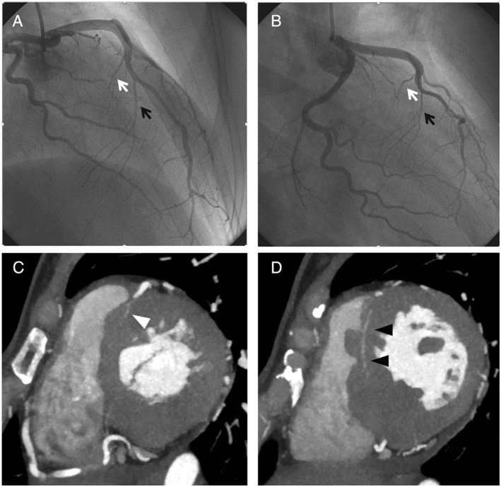
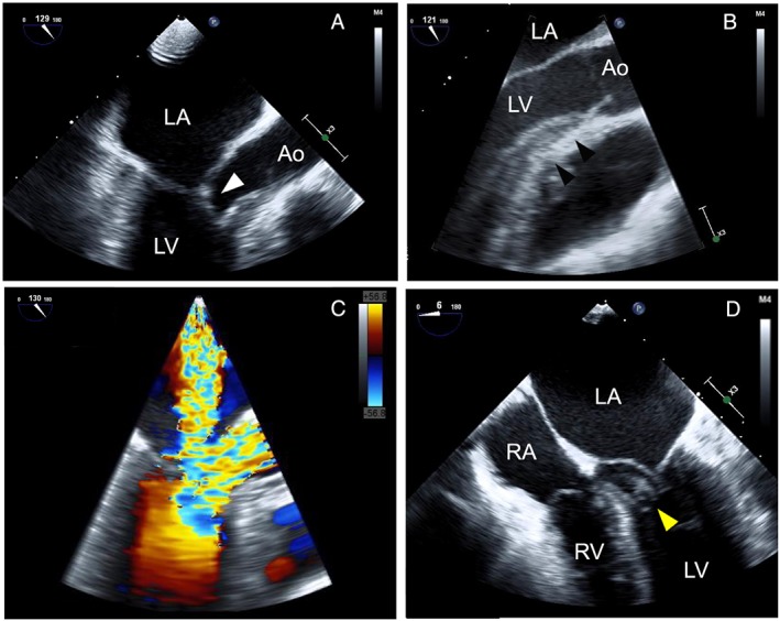
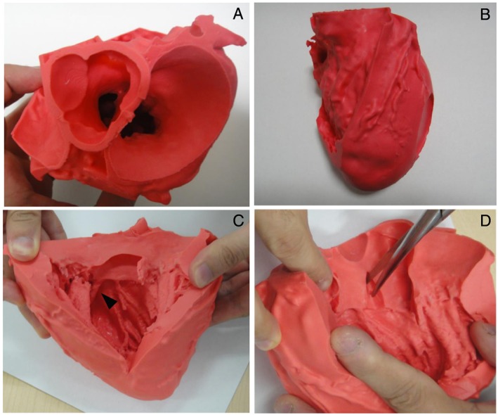
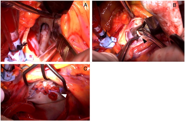
Both surgical myectomy and percutaneous transluminal septal myocardial ablation are effective treatments for drug-refractory symptomatic hypertrophic obstructive cardiomyopathy (HOCM). However, in some cases, it is not easy to elucidate the abnormal structure of left ventricular outflow obstruction to adopt these treatments. Here, we presented a young female patient with drug-refractory symptomatic HOCM. In this case, contrast-enhanced computed tomography enabled us to assess the suitability of percutaneous transluminal septal myocardial ablation. By creating three-dimensional printed models using computed tomography data, we could also visualize intracardiac structure and simulate the surgical procedure. A multimodality assessment strategy is useful for evaluating patients complicated with drug-refractory symptomatic HOCM.
Keywords: 3D printing; Hypertrophic obstructive cardiomyopathy; Surgical myectomy.
© 2017 The Authors. ESC Heart Failure published by John Wiley & Sons Ltd on behalf of the European Society of Cardiology.
Figures
Similar articles
-
A novel approach in the use of radiofrequency catheter ablation of septal hypertrophy in hypertrophic obstructive cardiomyopathy.Indian Heart J. 2016 Sep-Oct;68(5):618-623. doi: 10.1016/j.ihj.2016.02.007. Epub 2016 Apr 14. Indian Heart J. 2016. PMID: 27773399 Free PMC article.
-
Successful percutaneous transluminal septal myocardial ablation through a right superior septal artery for a patient with hypertrophic obstructive cardiomyopathy.Cardiovasc Interv Ther. 2015 Oct;30(4):377-81. doi: 10.1007/s12928-014-0310-4. Epub 2014 Dec 13. Cardiovasc Interv Ther. 2015. PMID: 25502014
-
Two-Port Thoracoscopic Myectomy for Hypertrophic Cardiomyopathy With Three-Dimensional Printing.Ann Thorac Surg. 2021 Mar;111(3):e165-e168. doi: 10.1016/j.athoracsur.2020.05.183. Epub 2020 Aug 7. Ann Thorac Surg. 2021. PMID: 32777215
-
Percutaneous transluminal septal myocardial ablation: past, present, and future.J Cardiol. 2022 Sep;80(3):211-217. doi: 10.1016/j.jjcc.2021.11.023. Epub 2021 Dec 16. J Cardiol. 2022. PMID: 34924238 Review.
-
Post-operative management of hypertrophic obstructive cardiomyopathy.Asian Cardiovasc Thorac Ann. 2022 Jan;30(1):57-63. doi: 10.1177/02184923211069189. Epub 2022 Feb 15. Asian Cardiovasc Thorac Ann. 2022. PMID: 35167344 Review.
Cited by
-
Three-dimensional printing to plan intracardiac operations.JTCVS Tech. 2021 Apr 20;9:101-108. doi: 10.1016/j.xjtc.2021.02.050. eCollection 2021 Oct. JTCVS Tech. 2021. PMID: 34647075 Free PMC article. No abstract available.
-
Multimodality imaging and three-dimensional printed model in patients with left ventricular outflow tract obstruction.ESC Heart Fail. 2020 Feb;7(1):320-324. doi: 10.1002/ehf2.12566. Epub 2019 Dec 11. ESC Heart Fail. 2020. PMID: 31825174 Free PMC article.
-
Three-dimensional printing for heart diseases: clinical application review.Biodes Manuf. 2021;4(3):675-687. doi: 10.1007/s42242-021-00125-8. Epub 2021 Apr 30. Biodes Manuf. 2021. PMID: 33948306 Free PMC article. Review.
-
Clinical situations for which 3D printing is considered an appropriate representation or extension of data contained in a medical imaging examination: adult cardiac conditions.3D Print Med. 2020 Sep 23;6(1):24. doi: 10.1186/s41205-020-00078-1. 3D Print Med. 2020. PMID: 32965536 Free PMC article.
-
The effects of septal myectomy and alcohol septal ablation for hypertrophic cardiomyopathy on the cardiac conduction system.J Interv Card Electrophysiol. 2018 Aug;52(3):403-408. doi: 10.1007/s10840-018-0433-0. Epub 2018 Aug 10. J Interv Card Electrophysiol. 2018. PMID: 30097789 Review.
References
-
- Elliott PM, Anastasakis A, Borger MA, Borggrefe M, Cecchi F, Charron P, Hagege AA, Lafont A, Limongelli G, Mahrholdt H, McKenna WJ, Mogensen J, Nihoyannopoulos P, Nistri S, Pieper PG, Pieske B, Rapezzi C, Rutten FH, Tillmanns C, Watkins H. 2014 ESC guidelines on diagnosis and management of hypertrophic cardiomyopathy: the task force for the diagnosis and management of hypertrophic cardiomyopathy of the European Society of Cardiology (ESC). Eur Heart J 2014; 35: 2733–2779. - PubMed
-
- Gersh BJ, Maron BJ, Bonow RO, Dearani JA, Fifer MA, Link MS, Naidu SS, Nishimura RA, Ommen SR, Rakowski H, Seidman CE, Towbin JA, Udelson JE, Yancy CW. 2011 ACCF/AHA guideline for the diagnosis and treatment of hypertrophic cardiomyopathy: executive summary: a report of the American College of Cardiology Foundation/American Heart Association Task Force on Practice Guidelines. Circulation 2011; 124: 2761–2796. - PubMed
-
- Maron BJ, Nishimura RA. Surgical septal myectomy versus alcohol septal ablation: assessing the status of the controversy in 2014. Circulation 2014; 130: 1617–1624. - PubMed
-
- Ghersin E, Soto V, Heldman AW. Multidetector computerized tomography can guide and document alcohol septal ablation in hypertrophic obstructive cardiomyopathy. Circulation 2011; 123: e5–e7. - PubMed
-
- Spratt JC, Langrish JP. Identifying the target septal perforator prior to alcohol septal ablation in hypertrophic obstructive cardiomyopathy: a new application for computed tomography coronary angiography. Heart 2011; 97: 1718–1719. - PubMed
Publication types
MeSH terms
Substances
LinkOut - more resources
Full Text Sources
Other Literature Sources
Medical
