

Non-aneurysmal subarachnoid hemorrhage: When is a second angiography indicated?
- PMID: 29154725
- PMCID: PMC5958500
- DOI: 10.1177/1971400917743100
Non-aneurysmal subarachnoid hemorrhage: When is a second angiography indicated?
Abstract
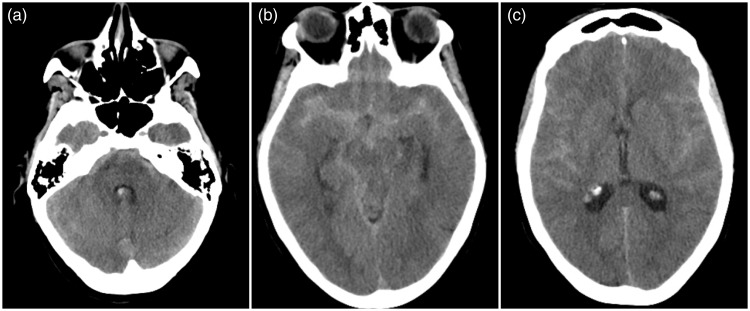
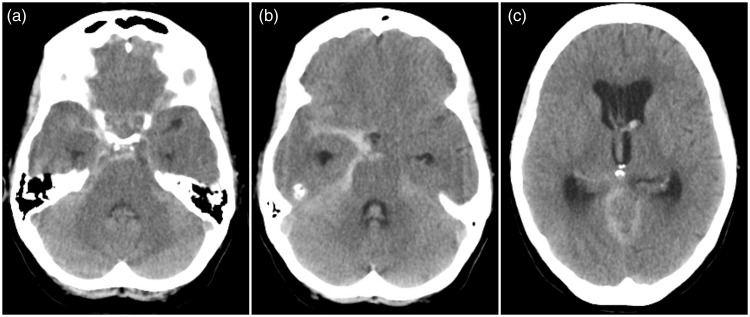
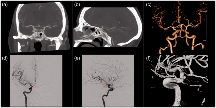
Purpose Repeat imaging in patients with non-aneurysmal subarachnoid hemorrhage (NASAH) remains controversial. We aim to report our experience with NASAH with different hemorrhage patterns, and to investigate the need for further diagnostic workup to determine the underlying cause of hemorrhage. Method We conducted a retrospective analysis of all spontaneous SAH with an initial negative computed tomography (CT) with angiography (CTA) and/or digital subtraction angiography (DSA) from October 2011 through May 2017. According to the bleeding pattern on the admission CT scan, NASAH was divided into two subgroups: (1) perimesencephalic SAH (PMSAH) and (2) non-perimesencephalic SAH (nPMSAH). Radiological data included the admission CT, CTA, DSA, and magnetic resonance imaging (MRI) with angiography (MRA). Results Seventy-four patients met the inclusion criteria. Thirty-nine (52.7%) patients had PMSAH on the initial CT scan, and 35 (47.3%) had nPMSAH. All underwent CTA and/or DSA revealing no vascular abnormalities. Forty-seven (63.5%) patients underwent subsequent diagnostic workup. DSA was performed in all patients at least once. No abnormalities were found on the repeat DSA or other radiological follow-up studies except in one (1.4%) patient with nPMSAH, in whom a follow-up DSA revealed a small saccular anterior choroidal artery aneurysm, considered to be the source of hemorrhage. Conclusion A repeat DSA may not be needed in case of PMSAH, if the initial negative DSA is technically adequate with absence of hematoma and vasospasm. In contrast, a follow-up DSA should be mandatory for confirming or excluding vascular pathology in case of nPMSAH in order to prevent rebleeding.
Keywords: Non-aneurysmal subarachnoid hemorrhage; cerebral angiography; digital subtraction angiography; perimesencephalic subarachnoid hemorrhage.
Figures

Comment in
-
Letter to the Editor regarding "non-aneurysmal subarachnoid hemorrhage: When is a second angiography indicated?".Neuroradiol J. 2018 Aug;31(4):449. doi: 10.1177/1971400918772166. Epub 2018 Apr 24. Neuroradiol J. 2018. PMID: 29687746 Free PMC article. No abstract available.
-
Reply to the letter to editor regarding: Non-aneurysmal subarachnoid hemorrhage: When is a second angiography indicated?Neuroradiol J. 2018 Aug;31(4):450-451. doi: 10.1177/1971400918778151. Epub 2018 May 17. Neuroradiol J. 2018. PMID: 29770724 Free PMC article. No abstract available.
References
-
- Rinkel GJ, Wijdicks EF, Vermeulen M, et al. The clinical course of perimesencephalic nonaneurysmal subarachnoid hemorrhage. Ann Neurol 1991; 29: 463–468. - PubMed
-
- Rinkel GJ, van Gijn J, Wijdicks EF. Subarachnoid hemorrhage without detectable aneurysm. A review of the causes. Stroke 1993; 24: 1403–1409. - PubMed
-
- van der Schaaf IC, Velthuis BK, Gouw A, et al. Venous drainage in perimesencephalic hemorrhage. Stroke 2004; 35: 1614–1618. - PubMed
-
- Rinkel GJ, Wijdicks EF, Hasan D, et al. Outcome in patients with subarachnoid haemorrhage and negative angiography according to pattern of haemorrhage on computed tomography. Lancet 1991; 338: 964–968. - PubMed
Publication types
MeSH terms
LinkOut - more resources
Full Text Sources
Other Literature Sources
Medical
Miscellaneous
