

Inflammatory Renin-Angiotensin System Disruption Attenuates Sensory Hyperinnervation and Mechanical Hypersensitivity in a Rat Model of Provoked Vestibulodynia
- PMID: 29155208
- PMCID: PMC5811351
- DOI: 10.1016/j.jpain.2017.10.006
Inflammatory Renin-Angiotensin System Disruption Attenuates Sensory Hyperinnervation and Mechanical Hypersensitivity in a Rat Model of Provoked Vestibulodynia
Abstract
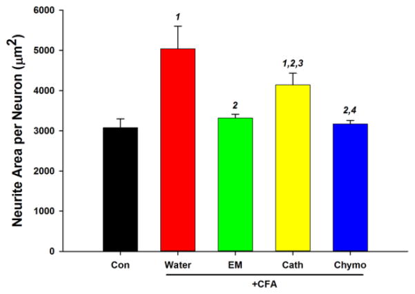
Vestibulodynia is characterized by perivaginal mechanical hypersensitivity, hyperinnervation, and abundant inflammatory cells expressing renin-angiotensin system proteins. We developed a tractable rat model of vestibulodynia to further assess the contributions of the renin-angiotensin system. Complete Freund's adjuvant injected into the posterior vestibule induced marked vestibular hypersensitivity throughout a 7-day test period. Numbers of axons immunoreactive for PGP9.5, calcitonin gene-related peptide, and GFRα2 were increased. Numbers of macrophages and T cells were also increased whereas B cells were not. Renin-angiotensin-associated proteins were abundant, with T cells as well as macrophages contributing to increased renin and angiotensinogen. Media conditioned with inflamed vestibular tissue promoted neurite sprouting by rat dorsal root ganglion neurons in vitro, and this was blocked by the angiotensin II receptor type 2 receptor antagonist PD123319 or by an angiotensin II function blocking antibody. Sensory axon sprouting induced by inflamed tissue was dependent on activity of angiotensin-converting enzyme or chymase, but not cathepsin G. Thus, vestibular Complete Freund's adjuvant injection substantially recapitulates changes seen in patients with provoked vestibulodynia, and shows that manipulation of the local inflammatory renin-angiotensin system may be a useful therapeutic strategy.
Perspective: This study provides evidence that inflammation of the rat vestibule induces a phenotype recapitulating behavioral and cytological features of human vestibulodynia. The model confirms a crucial role of the local inflammatory renin-angiotensin system in hypersensitivity and hyperinnervation. Targeting this system holds promise for developing new nonopioid analgesic treatment strategies.
Keywords: Allodynia; angiotensin II receptor type 2; axon sprouting; growth factors; hormones.
Copyright © 2017 The American Pain Society. Published by Elsevier Inc. All rights reserved.
Figures


Similar articles
-
A Local Inflammatory Renin-Angiotensin System Drives Sensory Axon Sprouting in Provoked Vestibulodynia.J Pain. 2017 May;18(5):511-525. doi: 10.1016/j.jpain.2016.12.008. Epub 2017 Jan 3. J Pain. 2017. PMID: 28062309 Free PMC article.
-
Angiotensin II receptor type 2 activation is required for cutaneous sensory hyperinnervation and hypersensitivity in a rat hind paw model of inflammatory pain.J Pain. 2013 Oct;14(10):1053-65. doi: 10.1016/j.jpain.2013.04.002. Epub 2013 May 30. J Pain. 2013. PMID: 23726047 Free PMC article.
-
Estrogen elicits dorsal root ganglion axon sprouting via a renin-angiotensin system.Endocrinology. 2008 Jul;149(7):3452-60. doi: 10.1210/en.2008-0061. Epub 2008 Apr 3. Endocrinology. 2008. PMID: 18388195 Free PMC article.
-
Targeting angiotensin II type 2 receptor pathways to treat neuropathic pain and inflammatory pain.Expert Opin Ther Targets. 2015 Jan;19(1):25-35. doi: 10.1517/14728222.2014.957673. Epub 2014 Oct 15. Expert Opin Ther Targets. 2015. PMID: 25315162 Review.
-
Innervation of the human vulvar vestibule: A comprehensive review.Clin Anat. 2023 Jan;36(1):18-27. doi: 10.1002/ca.23966. Epub 2022 Oct 19. Clin Anat. 2023. PMID: 36216779 Review.
Cited by
-
Angiotensin type 2 receptor antagonism as a new target to manage gout.Inflammopharmacology. 2022 Dec;30(6):2399-2410. doi: 10.1007/s10787-022-01076-x. Epub 2022 Sep 29. Inflammopharmacology. 2022. PMID: 36173505 Free PMC article.
-
Exploring Localized Provoked Vulvodynia: Insights from Animal Model Research.Int J Mol Sci. 2024 Apr 11;25(8):4261. doi: 10.3390/ijms25084261. Int J Mol Sci. 2024. PMID: 38673846 Free PMC article.
-
The Renin-Angiotensin System: The Challenge behind Autoimmune Dermatological Diseases.Diagnostics (Basel). 2023 Nov 7;13(22):3398. doi: 10.3390/diagnostics13223398. Diagnostics (Basel). 2023. PMID: 37998534 Free PMC article. Review.
-
Immune mechanisms in vulvodynia: key roles for mast cells and fibroblasts.Front Cell Infect Microbiol. 2023 Jun 8;13:1215380. doi: 10.3389/fcimb.2023.1215380. eCollection 2023. Front Cell Infect Microbiol. 2023. PMID: 37360527 Free PMC article. Review.
-
The Angiotensin II Type 2 Receptor, a Target for Protection and Regeneration of the Peripheral Nervous System?Pharmaceuticals (Basel). 2021 Feb 24;14(3):175. doi: 10.3390/ph14030175. Pharmaceuticals (Basel). 2021. PMID: 33668331 Free PMC article. Review.
References
-
- Alfredson H, Ohberg L, Forsgren S. Is vasculo-neural ingrowth the cause of pain in chronic Achilles tendinosis? An investigation using ultrasonography and colour Doppler, immunohistochemistry, and diagnostic injections. Knee Surg Sports Traumatol Arthrosc. 2003;11:334–338. - PubMed
-
- Anand U, Facer P, Yiangou Y, Sinisi M, Fox M, McCarthy T, Bountra C, Korchev YE, Anand P. Angiotensin II type 2 receptor (AT(2) R) localization and antagonist-mediated inhibition of capsaicin responses and neurite outgrowth in human and rat sensory neurons. Eur J Pain. 2013;17:1012–1026. - PMC - PubMed
-
- Anand U, Yiangou Y, Sinisi M, Fox M, MacQuillan A, Quick T, Korchev YE, Bountra C, McCarthy T, Anand P. Mechanisms underlying clinical efficacy of Angiotensin II type 2 receptor (AT2R) antagonist EMA401 in neuropathic pain: clinical tissue and in vitro studies. Molecular pain. 2015;11:38. - PMC - PubMed
Publication types
MeSH terms
Substances
Grants and funding
LinkOut - more resources
Full Text Sources
Other Literature Sources
