

A Novel Use of Three-dimensional High-frequency Ultrasonography for Early Pregnancy Characterization in the Mouse
- PMID: 29155779
- PMCID: PMC5695270
- DOI: 10.3791/56207
A Novel Use of Three-dimensional High-frequency Ultrasonography for Early Pregnancy Characterization in the Mouse
Abstract
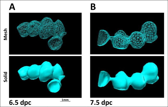
High-frequency ultrasonography (HFUS) is a common method to non-invasively monitor the real-time development of the human fetus in utero. The mouse is routinely used as an in vivo model to study embryo implantation and pregnancy progression. Unfortunately, such murine studies require pregnancy interruption to enable follow-up phenotypic analysis. To address this issue, we used three-dimensional (3-D) reconstruction of HFUS imaging data for early detection and characterization of murine embryo implantation sites and their individual developmental progression in utero. Combining HFUS imaging with 3-D reconstruction and modeling, we were able to accurately quantify embryo implantation site number as well as monitor developmental progression in pregnant C57BL6J/129S mice from 5.5 days post coitus (d.p.c.) through to 9.5 d.p.c. with the use of a transducer. Measurements included: number, location, and volume of implantation sites as well as inter-implantation site spacing; embryo viability was assessed by cardiac activity monitoring. In the immediate post-implantation period (5.5 to 8.5 d.p.c.), 3-D reconstruction of the gravid uterus in both mesh and solid overlay format enabled visual representation of the developing pregnancies within each uterine horn. As genetically engineered mice continue to be used to characterize female reproductive phenotypes derived from uterine dysfunction, this method offers a new approach to detect, quantify, and characterize early implantation events in vivo. This novel use of 3-D HFUS imaging demonstrates the ability to successfully detect, visualize, and characterize embryo-implantation sites during early murine pregnancy in a non-invasive manner. The technology offers a significant improvement over current methods, which rely on the interruption of pregnancies for gross tissue and histopathologic characterization. Here we use a video and text format to describe how to successfully perform ultrasounds of early murine pregnancy to generate reliable and reproducible data with reconstruction of the uterine form in mesh and solid 3-D images.


References
-
- Rai R, Regan L. Recurrent miscarriage. Lancet. 2006;368(9535):601–611. - PubMed
-
- Sugiura-Ogasawara M, Ozaki Y, Suzumori N. Management of recurrent miscarriage. J Obstet Gynaecol Res. 2014;40(5):1174–1179. - PubMed
-
- Kutteh WH. Novel strategies for the management of recurrent pregnancy loss. Semin Reprod Med. 2015;33(3):161–168. - PubMed
-
- Page JM, Silver RM. Genetic Causes of Recurrent Pregnancy Loss. Clin Obstet Gynecol. 2016;59(3):498–508. - PubMed
Publication types
MeSH terms
Grants and funding
LinkOut - more resources
Full Text Sources
Other Literature Sources
Research Materials
