. 2017 Nov 20;13(11):e1005799.
doi: 10.1371/journal.pcbi.1005799. eCollection 2017 Nov.

Multivariate pattern dependence

Affiliations

Multivariate pattern dependence

Stefano Anzellotti et al. PLoS Comput Biol. .

Abstract

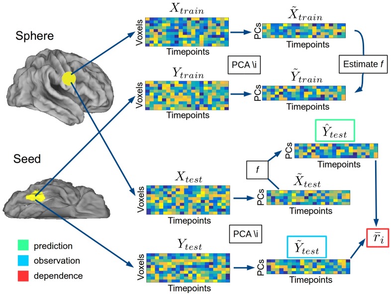
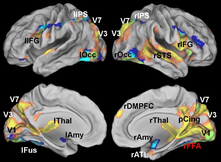
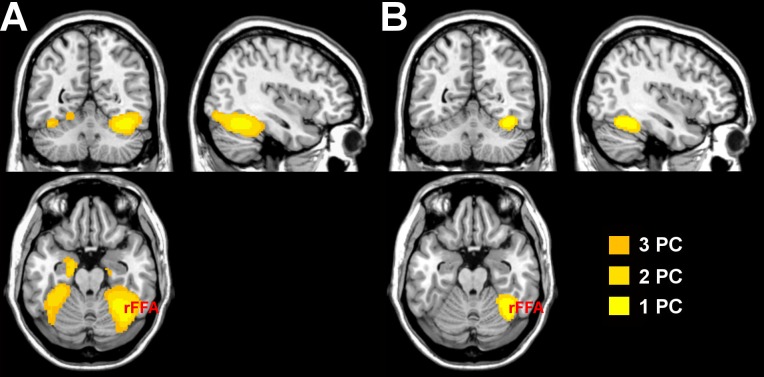
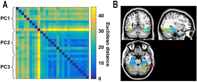
When we perform a cognitive task, multiple brain regions are engaged. Understanding how these regions interact is a fundamental step to uncover the neural bases of behavior. Most research on the interactions between brain regions has focused on the univariate responses in the regions. However, fine grained patterns of response encode important information, as shown by multivariate pattern analysis. In the present article, we introduce and apply multivariate pattern dependence (MVPD): a technique to study the statistical dependence between brain regions in humans in terms of the multivariate relations between their patterns of responses. MVPD characterizes the responses in each brain region as trajectories in region-specific multidimensional spaces, and models the multivariate relationship between these trajectories. We applied MVPD to the posterior superior temporal sulcus (pSTS) and to the fusiform face area (FFA), using a searchlight approach to reveal interactions between these seed regions and the rest of the brain. Across two different experiments, MVPD identified significant statistical dependence not detected by standard functional connectivity. Additionally, MVPD outperformed univariate connectivity in its ability to explain independent variance in the responses of individual voxels. In the end, MVPD uncovered different connectivity profiles associated with different representational subspaces of FFA: the first principal component of FFA shows differential connectivity with occipital and parietal regions implicated in the processing of low-level properties of faces, while the second and third components show differential connectivity with anterior temporal regions implicated in the processing of invariant representations of face identity.


Conflict of interest statement

The authors have declared that no competing interests exist.

Figures

Fig 1
Fig 1. Analysis pipeline.
Fig 2
Fig 2. Brain regions showing statistical dependence with the right pSTS as identified by standard functional connectivity (blue) and multivariate pattern dependence (MVPD, yellow) at a voxelwise FWE-corrected threshold p < 0.05.
MVPD additionally identified statistical dependence with the posterior cingulate, and with larger portions of posterior STS bilaterally.
Fig 3
Fig 3
A) Comparison between statistical dependence measured with standard functional connectivity (‘univariate dependence’, blue), univariate dependence with leave-one-out predictions (red), and multivariate dependence with leave-one-out predictions (MVPD, yellow) at a voxelwise FWE-corrected threshold p < 0.05. Predicting independent data is a more stringent test of dependence: univariate dependence with leave-one-out predictions individuates fewer significant voxels than standard functional connectivity. Despite the stringent criterion imposed by independent predictions, MVPD with leave-one-out predictions outperforms both univariate dependence methods, identifying significant statistical dependence with regions of posterior cingulate and a broader extent of the superior temporal sulcus. B) Cross-validated R-squared with MVPD, thresholded at 20%.
Fig 4
Fig 4. MVPD calculated after removing the univariate signal, in coronal (A), axial (B), and sagittal left (C) and right (D) views.
In this analysis, the average timecourse in the seed and each sphere is zero, and only the patterns of responses are left. As a consequence, this analysis is fully complementary to standard functional connectivity. Even after removing the univariate signal, MVPD detected significant statistical dependence between the right pSTS and posterior cingulate as well as the left STS.
Fig 5
Fig 5. Brain regions showing statistical dependence with the FFA as identified by standard functional connectivity (blue) and multivariate pattern connectivity (MVPD, yellow) at a voxelwise FWE-corrected threshold p < 0.05.
MVPD, but not standard functional connectivity, identified statistical dependence with regions of posterior cingulate, the right superior temporal sulcus, the right anterior temporal lobe, the right DMPFC and regions of the dorsal visual stream. Standard functional connectivity identified statistical dependence with the amygdala that was not detected by MVPD.
Fig 6
Fig 6
A) Regions with cross-validated R-squared above 5%, as measured by MVPD with 1, 2 and 3 principal components. B) Regions with cross-validated R-squared above 10%, as measured by MVPD with 1, 2 and 3 principal components.
Fig 7
Fig 7
A) Similarity matrix between the whole-brain maps of r values obtained with MVPD for each participant reflecting the statistical dependence between each voxel and the first, second, and third PC respectively in the FFA seed. B) Top 5000 voxels showing highest r values for the first PC (in yellow), and for the second and third PCs (in blue). Different subspaces of FFA responses show different MVPD profiles, with the first dimension showing greatest statistical dependence with posterior ventral temporal regions and regions in the dorsal visual stream, and the second and third dimensions showing greatest statistical dependence with anterior temporal regions.

Similar articles

Cited by

References

    1. Anzellotti Stefano and Caramazza Alfonso From parts to identity: invariance and sensitivity of face representations to different face halves, Cerebral Cortex 2015. 26(5):1900–1909. doi: 10.1093/cercor/bhu337 - DOI - PubMed
    1. Fedorenko Evelina and Thompson-Schill Sharon L Reworking the language network, Trends in cognitive sciences 2014. 18(3):120–126. doi: 10.1016/j.tics.2013.12.006 - DOI - PMC - PubMed
    1. Ishai Alumit Let’s face it: it’s a cortical network, Neuroimage 2008. 40(2):415–419. doi: 10.1016/j.neuroimage.2007.10.040 - DOI - PubMed
    1. Gallagher Helen L and Frith Christopher D Functional imaging of ‘theory of mind’, Trends in cognitive sciences 2003. 7(2):77–83. doi: 10.1016/S1364-6613(02)00025-6 - DOI - PubMed
    1. Le Bihan Denis and Mangin Jean-François and Poupon Cyril and Clark Chris A and Pappata Sabina and Molko Nicolas and Chabriat Hughes Diffusion tensor imaging: concepts and applications, Journal of magnetic resonance imaging 2001. 13(4):534–546. doi: 10.1002/jmri.1076 - DOI - PubMed