Case Reports
. 2018 Jan 1;77(1):50-63.
doi: 10.1093/jnen/nlx101.

Dementia After Moderate-Severe Traumatic Brain Injury: Coexistence of Multiple Proteinopathies

Affiliations
Case Reports

Dementia After Moderate-Severe Traumatic Brain Injury: Coexistence of Multiple Proteinopathies

Kimbra Kenney et al. J Neuropathol Exp Neurol. .

Abstract

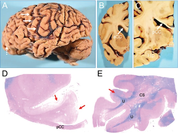
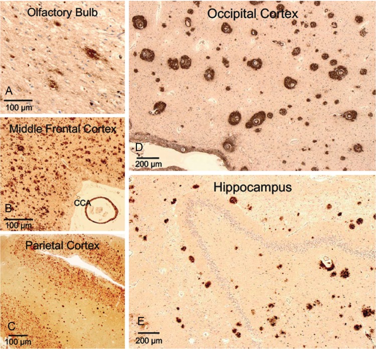
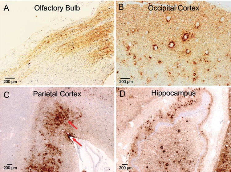
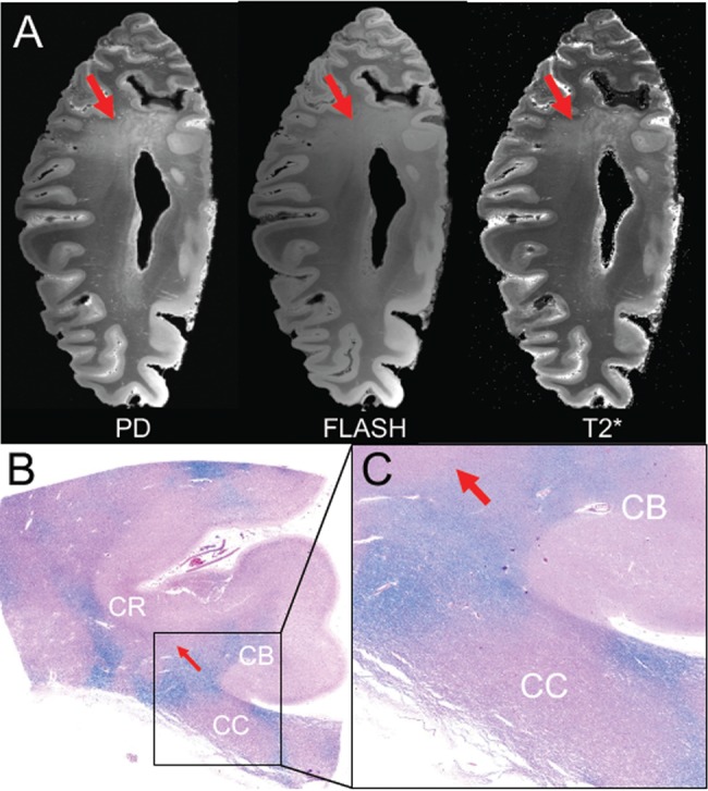
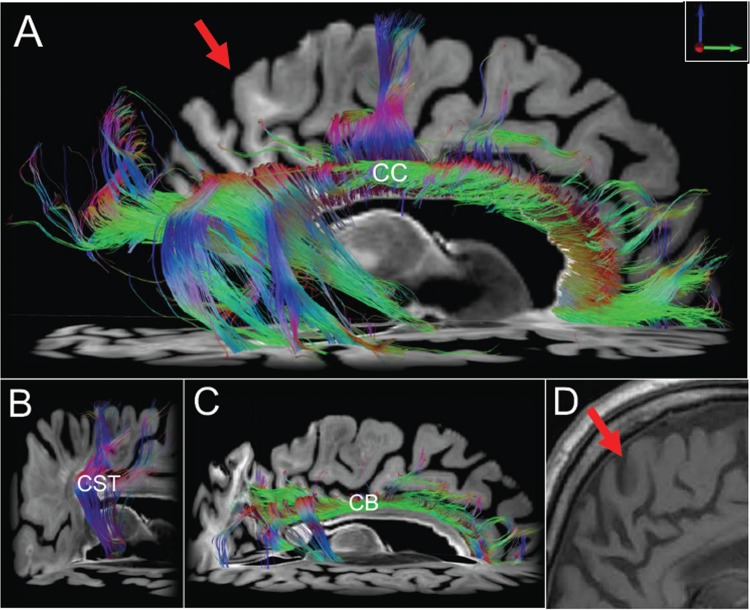
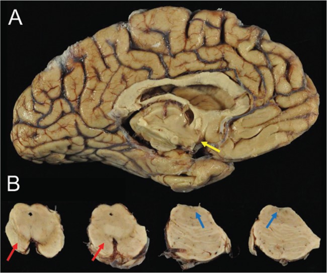
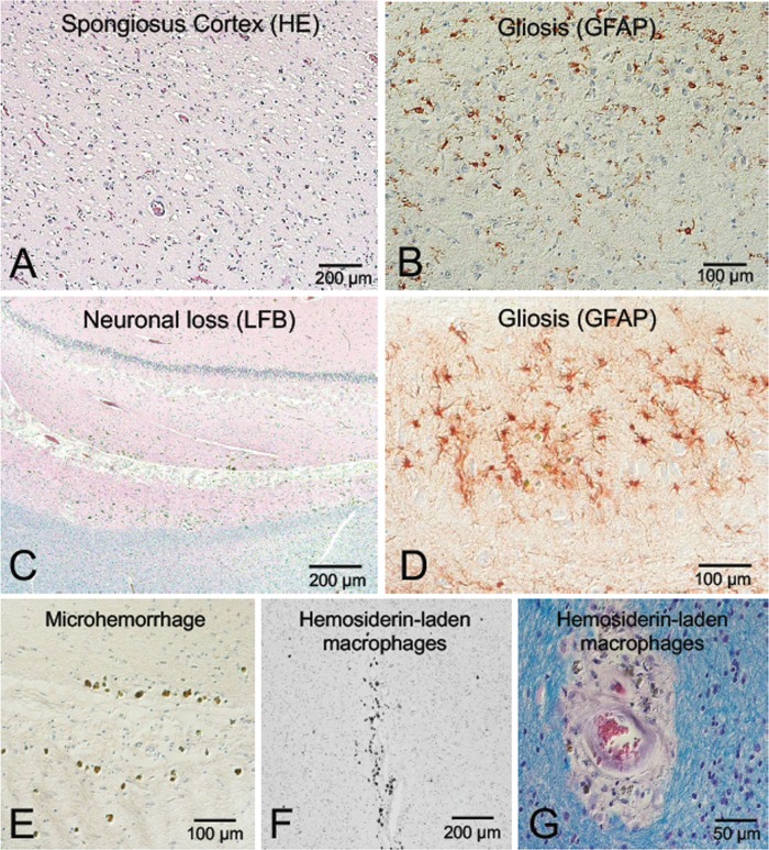
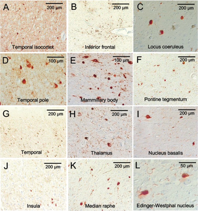
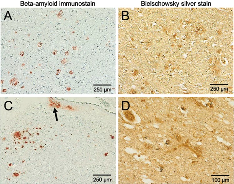
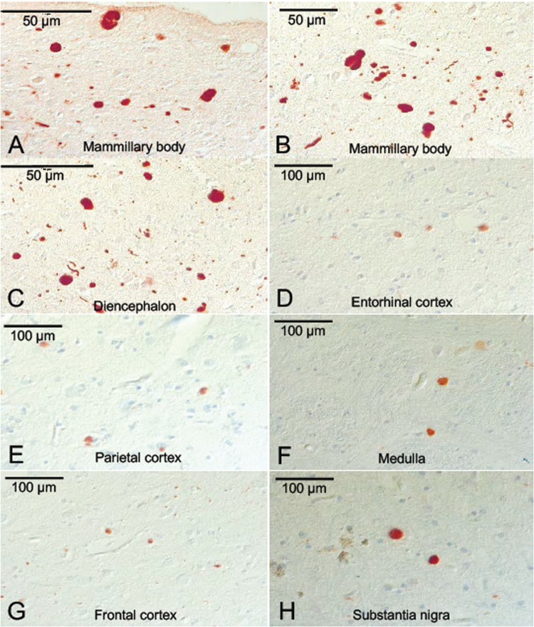
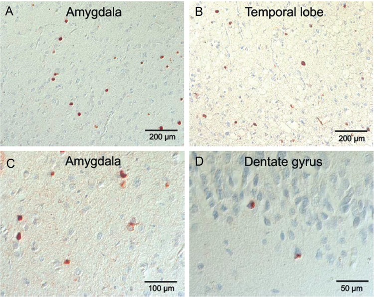
We report the clinical, neuroimaging, and neuropathologic characteristics of 2 patients who developed early onset dementia after a moderate-severe traumatic brain injury (TBI). Neuropathological evaluation revealed abundant β-amyloid neuritic and cored plaques, diffuse β-amyloid plaques, and frequent hyperphosphorylated-tau neurofibrillary tangles (NFT) involving much of the cortex, including insula and mammillary bodies in both cases. Case 1 additionally showed NFTs in both the superficial and deep cortical layers, occasional perivascular and depth-of-sulci NFTs, and parietal white matter rarefaction, which corresponded with decreased parietal fiber tracts observed on ex vivo MRI. Case 2 additionally showed NFT predominance in the superficial layers of the cortex, hypothalamus and brainstem, diffuse Lewy bodies in the cortex, amygdala and brainstem, and intraneuronal TDP-43 inclusions. The neuropathologic diagnoses were atypical Alzheimer disease (AD) with features of chronic traumatic encephalopathy and white matter loss (Case 1), and atypical AD, dementia with Lewy bodies and coexistent TDP-43 pathology (Case 2). These findings support an epidemiological association between TBI and dementia and further characterize the variety of misfolded proteins that may accumulate after TBI. Analyses with comprehensive clinical, imaging, genetic, and neuropathological data are required to characterize the full clinicopathological spectrum associated with dementias occurring after moderate-severe TBI.

Keywords: Hyperphosphorylated tau; Neurodegeneration; Neurofibrillary tangle; Proteinopathy; Traumatic brain injury; α-Synuclein; β-Amyloid.


Figures

FIGURE 1.
FIGURE 1.
Macroscopic appearance of the brain of a subject (Case 1) with dementia after a moderate-severe TBI. Thinning of the posterior corpus callosum and myelin loss. Gross appearance of brain specimen from Case 1. (A) The intact brain, left cerebral (contusion labeled with dashed arrows) and cerebellar hemispheres. (B) Coronal cross-section of the left hemisphere at the level of the caudate/putamen and anterior corpus callosum (aCC). (C) Coronal large cross-section of left hemisphere at the level of the hippocampus (Hip) and posterior corpus callosum (pCC). Of note, the marked intrasulcal space enlargement (as per diffuse cortical atrophy) (arrows, A) and marked thinning of the posterior corpus callosum (pCC) (arrow, C) contrasts the relatively preserved thickness of the anterior corpus callosum (aCC) (arrow, B), and the relatively normal appearance of the hippocampus (C). Panel C shows a LFB-hematoxylin stain of the left posterior corpus callosum (pCC). Panel D shows a LFB-hematoxylin stain of the left superior temporal cortex. These stains demonstrate extensive loss of myelin in the pCC and adjacent centrum semiovale (CS). Of note, the patchy myelin loss in temporal lobe spares the U-fibers (U). Cortical pathology associated with contusions are indicated by red arrows in panels C and D. The images were obtained using a 2.5× objective.
FIGURE 2.
FIGURE 2.
Extracellular β-amyloid accumulation in the cortex of a subject (Case 1) with dementia after moderate-severe TBI. High levels of extracellular β-amyloid accumulation detected by 4G8 antibody (1–42 β-amyloid) across various regions of the brain: olfactory bulb (A), middle frontal cortex (B), parietal cortex (C), occipital cortex (D), and hippocampus (E). Other notable findings include β-amyloid-positive cerebral congophilic angiopathy (CCA) in the vessels of the middle frontal cortex (B) and high frequency of diffuse β-amyloid plaques in the middle frontal cortex (B), parietal cortex (C), occipital cortex (D), and hippocampus (E). In the occipital cortex (D), immunohistochemistry (4G8) shows peculiar and unusually dense accumulation of β-amyloid around intracortical vessels. Objective 20 × (olfactory bulb; A), 5 × (middle frontal [B], parietal [C], occipital [D] cortices, and hippocampus [E]).
FIGURE 3.
FIGURE 3.
AD- and CTE-like p-tau accumulation in the cortex of a subject (Case 1) with dementia after moderate-severe TBI. High levels of p-tau accumulation were seen across different regions of the brain: olfactory bulb, parietal cortex, occipital cortex, and hippocampus. Importantly, we observed the coexistence of 2 different types of p-tau pathology: AD-like and CTE-like p-tau pathology (red arrows, parietal cortex). This is a rare finding that is difficult to identify when diffuse and high levels of p-tau deposition are present in a brain. Tau immunohistochemistry with AT8 antibody. Objective 5 × (olfactory bulb, occipital cortex); 2.5 × (parietal cortex, hippocampus). (A) A portion of the olfactory bulb and tract positive for p-tau lesions; (B) p-tau lesions in the occipital cortex (note the perivascular distribution of p-tau pathology); (C) rarely observed copresence of 2 types of p-tau accumulation in the same region (parietal cortex): AD pathology (across all deeper cortical layers) and p-tau sulcal pathology (more superficial cortical layers), which is pathognomonic of CTE; (D) Diffuse p-tau pathology across all hippocampus.
FIGURE 4.
FIGURE 4.
Imaging-histopathological correlations in a subject (Case 1) with dementia after a moderate-severe TBI. (A) Axial images from the proton density (PD) map, FLASH acquisition and T2* parameter map (T2*) derived from the ex vivo 7T MRI scan demonstrate rarification of myelin in the parietal white matter (red arrows). (B) Corresponding section from the parietal region that underwent ex vivo MRI. The histological section was stained with hematoxylin and eosin (H&E) and counter-stained with Luxol fast blue (LFB) for detection of myelin. This histological section (B), which is shown in zoomed view in panel (C), reveals patches of myelin loss throughout the coronal radiata (CR) and corpus callosum (CC). The LFB stain is more prominent in the cingulum bundle (CB), indicating that this white matter pathway is relatively spared.
FIGURE 5.
FIGURE 5.
Diffusion tractography findings of white matter in a subject (Case 1) with dementia after a moderate-severe TBI. Diffusion tractography results indicate focal regions of white matter injury. (A) Right lateral view of interhemispheric, transcallosal fiber tracts generated with a region of interest traced in the corpus callosum (CC). Fiber tracts from nearby white matter bundles (e.g., fornix, cingulum bundle) have been eliminated to optimize visualization. Transcallosal tracts are intact in parts of the frontal and temporal lobes, but they are disrupted in other regions such as the parietal lobe (arrow). These parietal tractography findings are consistent with the ex vivo 7T images seen in Figure 7A that show rarification of the parietal hemispheric white matter, as well as the histopathologic data in Figures 7B and C showing white matter injury in this region. To exclude the possibility that global loss of white matter and/or technical artifact is responsible for the transcallosal tractography results, we show relative preservation of corticospinal tract (CST) fibers from a right lateral view (B) and cingulum bundle (CB) fibers from a right lateral view (C). Corresponding imaging from an MRI (D) obtained 4 years before his death does not show the extensive gliosis visible in the ex vivo imaging (arrow).
FIGURE 6.
FIGURE 6.
Macroscopic appearance of the brain of a subject (Case 2) with dementia after a single severe TBI. (A) There was marked atrophy of cerebral cortex, corpus callosum, and medial temporal lobe with severe neuronal loss in amygdala, mammillary bodies (yellow arrow), entorhinal cortex, and hypothalamus. (B) Images show (from left to right) mesencephalon including substantia nigra (SN) and locus coeruleus (LC) with pigmented neuronal loss in SN (red arrows) and LC (blue arrows).
FIGURE 7.
FIGURE 7.
Neuronal loss, spongiform change, vascular lesions, and hemosiderin-laden macrophages in a subject (Case 2) with dementia after a single severe TBI. (A, B) Images showing temporal cortex neuronal loss and spongiosus cortex; 20× magnification with hematoxylin and eosin (H&E) staining (A) and GFAP immunohistochemistry (B). (C, D) Images showing neuronal loss and gliosis in hippocampus; 10× magnification of CA1 region stained with H&E and Luxol-fast blue (LFB) myelin staining (C), and 40× magnification of GFAP immunohistochemistry (D). (E–G) Images showing microhemorrhage in the hippocampus and white matter with perivascular hemosiderin-laden macrophages; 20× magnification with H&E of CA1 region of hippocampus (E); 10× magnification with H&E of white matter (F); and 40× magnification with LFB-H&E of white matter (G).
FIGURE 8.
FIGURE 8.
p-Tau accumulation in the brain of a subject (Case 2) with dementia after a single severe TBI. Atypical pattern and distribution of p-tau neurofibrillary pathology, predominantly involving superficial layers of cerebral cortices (B), with dotlike neurites (A, B, D, G, J), but also throughout midbrain, deep gray and brainstem nuclei (C, E, F, H, I, K, L). Immunostaining for p-tau with AT8 histochemistry at 10×–40× magnification.
FIGURE 9.
FIGURE 9.
β-Amyloid accumulation in the cortex and leptomeninges of a subject (Case 2) with dementia after a severe TBI. Sparse Aβ-plaques, predominantly diffuse plaques with vascular amyloid deposits in the leptomeninges (C, arrow). β-Amyloid immunostain (A, C) and Bielschowsky silver stain (B, D), 10× magnification.
FIGURE 10.
FIGURE 10.
α-Synuclein pathology in the brain of a subject (Case 2) with dementia after a severe TBI. α-Synuclein-positive Lewy bodies and Lewy neurites in entorhinal, parietal and frontal cortices, mammillary body, diencephalon and substantia nigra (SN), medial temporal cortex (A–H) with unusually large, voluminous α-synuclein-positive axonal spheroids in mammillary bodies, SN, medulla and diencephalon (A, B, C, F, H). Immunostaining with anti-phosphorylated α-synuclein at 20×–40× magnification.
FIGURE 11.
FIGURE 11.
TDP43 pathology in the cerebral cortex of a subject (Case 2) with dementia after a severe TBI. (A–D) TDP-43-positive inclusions in temporal lobe, amygdala and hippocampus, including dentate gyrus. Immunostaining with anti-TDP-43 antibody at 20×–40× magnification.

References

    1. Plassman BL, Havlik RJ, Steffens DC, et al.Documented head injury in early adulthood and risk of Alzheimer’s disease and other dementias. Neurology 2000; 55:1158–66 - PubMed
    1. Himanen L, Portin R, Isoniemi H, et al.Longitudinal cognitive changes in traumatic brain injury: a 30-year follow-up study. Neurology 2006; 66:187–9210.1212/01.wnl.0000194264.60150.d3 - DOI - PubMed
    1. Fleminger S, Oliver DL, Lovestone S, et al.Head injury as a risk factor for Alzheimer’s disease: the evidence 10 years on; a partial replication. J Neurol Neurosurg Psychiatry 2003; 74:857–62 - PMC - PubMed
    1. O'Meara ES, Kukull WA, Sheppard L, et al.Head injury and risk of Alzheimer’s disease by apolipoprotein E genotype. Am J Epidemiol 1997; 146:373–84 - PubMed
    1. Mehta KM, Ott A, Kalmijn S, et al.Head trauma and risk of dementia and Alzheimer’s disease: the Rotterdam study. Neurology 1999; 53:1959–62 - PubMed

Publication types

MeSH terms