

Site-Directed Mutagenesis of the Fibronectin Domains in Insulin Receptor-Related Receptor
- PMID: 29156593
- PMCID: PMC5713427
- DOI: 10.3390/ijms18112461
Site-Directed Mutagenesis of the Fibronectin Domains in Insulin Receptor-Related Receptor
Abstract
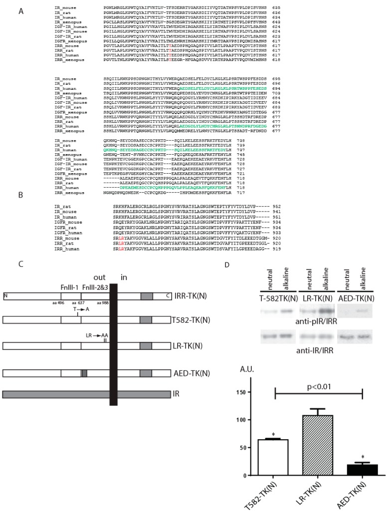
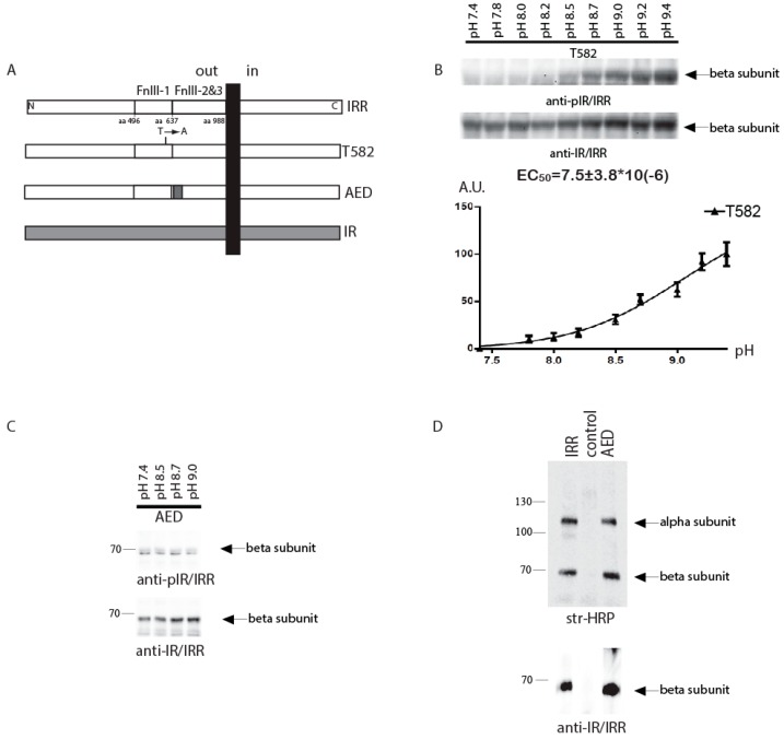
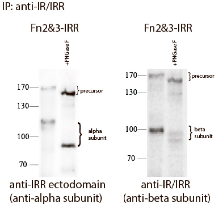
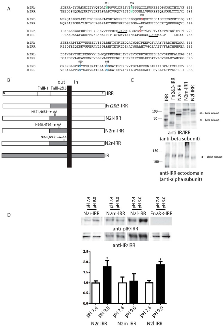
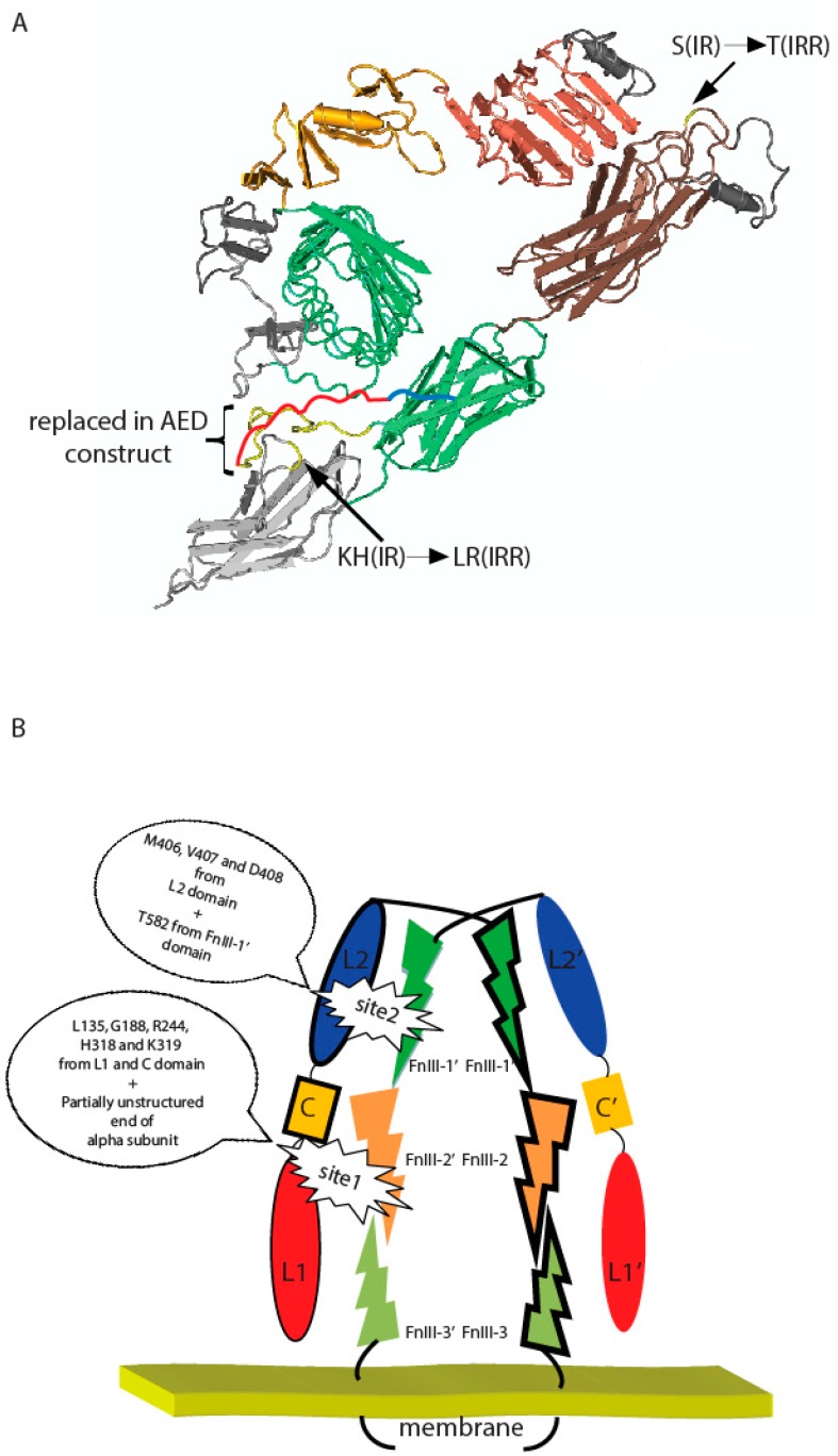
The orphan insulin receptor-related receptor (IRR), in contrast to its close homologs, the insulin receptor (IR) and insulin-like growth factor receptor (IGF-IR) can be activated by mildly alkaline extracellular medium. We have previously demonstrated that IRR activation is defined by its extracellular region, involves multiple domains, and shows positive cooperativity with two synergistic sites. By the analyses of point mutants and chimeras of IRR with IR in, we now address the role of the fibronectin type III (FnIII) repeats in the IRR pH-sensing. The first activation site includes the intrinsically disordered subdomain ID (646-716) within the FnIII-2 domain at the C-terminus of IRR alpha subunit together with closely located residues L135, G188, R244, H318, and K319 of L1 and C domains of the second subunit. The second site involves residue T582 of FnIII-1 domain at the top of IRR lambda-shape pyramid together with M406, V407, and D408 from L2 domain within the second subunit. A possible importance of the IRR carbohydrate moiety for its activation was also assessed. IRR is normally less glycosylated than IR and IGF-IR. Swapping both FnIII-2 and FnIII-3 IRR domains with those of IR shifted beta-subunit mass from 68 kDa for IRR to about 100 kDa due to increased glycosylation and abolished the IRR pH response. However, mutations of four asparagine residues, potential glycosylation sites in chimera IRR with swapped FnIII-2/3 domains of IR, decreased the chimera glycosylation and resulted in a partial restoration of IRR pH-sensing activity, suggesting that the extensive glycosylation of FnIII-2/3 provides steric hindrance for the alkali-induced rearrangement of the IRR ectodomain.
Keywords: alkaline medium; insulin receptor-related receptor; pH sensor; protein glycosylation; receptor tyrosine kinase activation; site directed mutagenesis; tyrosine phosphorylation.
Conflict of interest statement
The authors declare no conflict of interest.
Figures
Similar articles
-
Mapping of alkali-sensing sites of the insulin receptor-related receptor. The role of L2 and fibronectin domains.Biochimie. 2015 Apr;111:1-9. doi: 10.1016/j.biochi.2014.12.014. Epub 2015 Jan 15. Biochimie. 2015. PMID: 25597417
-
Structural determinants of the insulin receptor-related receptor activation by alkali.J Biol Chem. 2013 Nov 22;288(47):33884-33893. doi: 10.1074/jbc.M113.483172. Epub 2013 Oct 11. J Biol Chem. 2013. PMID: 24121506 Free PMC article.
-
A third fibronectin type III domain in the extracellular region of the insulin receptor family.FEBS Lett. 1998 Dec 18;441(2):331-6. doi: 10.1016/s0014-5793(98)01509-9. FEBS Lett. 1998. PMID: 9883910
-
Structural insights into ligand-induced activation of the insulin receptor.Acta Physiol (Oxf). 2008 Jan;192(1):3-9. doi: 10.1111/j.1748-1716.2007.01781.x. Acta Physiol (Oxf). 2008. PMID: 18171424 Review.
-
Insulin receptor-related receptor as an extracellular pH sensor involved in the regulation of acid-base balance.Biochim Biophys Acta. 2013 Oct;1834(10):2170-5. doi: 10.1016/j.bbapap.2012.11.011. Epub 2012 Dec 7. Biochim Biophys Acta. 2013. PMID: 23220417 Review.
Cited by
-
The dimeric ectodomain of the alkali-sensing insulin receptor-related receptor (ectoIRR) has a droplike shape.J Biol Chem. 2019 Nov 22;294(47):17790-17798. doi: 10.1074/jbc.RA119.010390. Epub 2019 Oct 15. J Biol Chem. 2019. PMID: 31615897 Free PMC article.
-
Probing Structure and Function of Alkali Sensor IRR with Monoclonal Antibodies.Biomolecules. 2020 Jul 16;10(7):1060. doi: 10.3390/biom10071060. Biomolecules. 2020. PMID: 32708676 Free PMC article.
References
-
- Shier P., Watt V.M. Primary structure of a putative receptor for a ligand of the insulin family. J. Biol. Chem. 1989;264:14605–14608. - PubMed
-
- Ullrich A., Gray A., Tam A.W., Yang-Feng T., Tsubokawa M., Collins C., Henzel W., Le Bon T., Kathuria S., Chen E., et al. Insulin-like growth factor I receptor primary structure: Comparison with insulin receptor suggests structural determinants that define functional specificity. EMBO J. 1986;5:2503–2512. - PMC - PubMed
MeSH terms
Substances
LinkOut - more resources
Full Text Sources
Other Literature Sources
Molecular Biology Databases
Research Materials
