. 2017 Nov 20;18(11):2473.
doi: 10.3390/ijms18112473.

Akt1 Stimulates Homologous Recombination Repair of DNA Double-Strand Breaks in a Rad51-Dependent Manner

Affiliations

Akt1 Stimulates Homologous Recombination Repair of DNA Double-Strand Breaks in a Rad51-Dependent Manner

Katharina Mueck et al. Int J Mol Sci. .

Abstract

Akt1 is known to promote non-homologous end-joining (NHEJ)-mediated DNA double-strand break (DSB) repair by stimulation of DNA-PKcs. In the present study, we investigated the effect of Akt1 on homologous recombination (HR)-dependent repair of radiation-induced DSBs in non-small cell lung cancer (NSCLC) cells A549 and H460. Akt1-knockdown (Akt1-KD) significantly reduced Rad51 protein level, Rad51 foci formation and its colocalization with γH2AX foci after irradiation. Moreover, Akt1-KD decreased clonogenicity after treatment with Mitomycin C and HR repair, as tested by an HR-reporter assay. Double knockdown of Akt1 and Rad51 did not lead to a further decrease in HR compared to the single knockdown of Rad51. Consequently, Akt1-KD significantly increased the number of residual DSBs after irradiation partially independent of the kinase activity of DNA-PKcs. Likewise, the number of residual BRCA1 foci, indicating unsuccessful HR events, also significantly increased in the irradiated cells after Akt1-KD. Together, the results of the study indicate that Akt1 seems to be a regulatory component in the HR repair of DSBs in a Rad51-dependent manner. Thus, based on this novel role of Akt1 in HR and the previously described role of Akt1 in NHEJ, we propose that targeting Akt1 could be an effective approach to selectively improve the killing of tumor cells by DSB-inducing cytotoxic agents, such as ionizing radiation.

Keywords: Akt1; DNA double-strand break repair; Rad51; homologous recombination; non-small cell lung cancer.


Conflict of interest statement

The authors declare no conflict of interest.

Figures
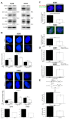
Figure 1
Figure 1
Akt1 promotes HR-dependent DSB repair. A549 and H460 cells were transfected with AKT1-siRNA or con-siRNA. (A) The protein levels of Akt1, Akt2 and Akt3 were analyzed by Western blotting. β-Actin was used as the loading control. The protein levels were normalized to those in the con-siRNA transfected cells. The data represent the mean ± SEM of the indicated number of independent experiments; (B) the Rad51 foci assay was performed as described in the Methods section at the indicated time points after irradiation. The bars represent the mean number of foci/cell ± SEM from at least 3 independent experiments. At least 276 nuclei per condition were evaluated. Bars showing the percentage of cells with at least 2 Rad51 foci/nucleus are based on data for the 8 h time point. Akt1-KD significantly decreased Rad51 foci formation (* p < 0.05, ** p < 0.01, *** p < 0.001, Student’s t-test); (C) 8 h after irradiation, the fraction of γH2AX foci colocalized with Rad51 foci was examined. The results are based on the mean number of foci from two independent experiments, a total of at least 175 nuclei per condition for A549 cells, and 2 independent experiments, a total of at least 197 counted nuclei for H460 cells (p < 0.001, Student’s t-test); (D) clonogenic survival after MMC treatment was analyzed (A549, n = 4, 12 data points; H460, n = 1, 3 data points; * p < 0.05, Student’s t-test); (E) HR-reporter assay was performed. A549 cells were treated with the indicated siRNAs or MK2206 (10 μM)/DMSO. The fraction of con-siRNA transfected cells, which were GFP-positive, equaled 0.56 ± 0.11%, the proportion of Akt1-KD cells equaled 0.40 ± 0.07%. The proportion of DMSO control cells, which were GFP-positive, equaled 1.52 ± 0.18%. The portion of GFP-positive MK2206 treated cells equaled 0.86 ± 0.10% (raw data). The raw values of GFP-positive cells were normalized to those in the con-siRNA/DMSO treated cells. Akt1-KD significantly reduced the fraction of GFP-positive cells (n = 3, 9 data points; ** p < 0.01, Student’s t-test). MK2206 treatment also significantly reduced the fraction of GFP-positive cells (n = 2, 3 data points; ** p < 0.01, Student’s t-test). PE: Plating Efficiency.
Figure 2
Figure 2
Stimulation of HR repair by Akt1 is dependent on Rad51. (A) HR-reporter assay was performed in A549 cells after transfection with the indicated siRNAs. The fraction of con-siRNA transfected cells, which were GFP-positive, equaled 0.46 ± 0.10%, for Akt1-KD 0.33 ± 0.05%, for Rad51-KD 0.24 ± 0.05%, for Akt1-KD+Rad51-KD 0.20 ± 0.03% (raw data). The raw values of GFP-positive cells were normalized to con-siRNA transfected cells. Asterisks indicate significant reduction of GFP-positive cells by Akt1-KD and Rad51-KD (n = 4, at least 8 data points; * p < 0.05, *** p < 0.001); (B) following transfection with AKT1-siRNA, A549 cells were irradiated, and 8 h later, the cytoplasmic and nuclear fractions were prepared. Rad51 protein levels were determined by Western blotting. GAPDH and Lamin A/C were used as cytoplasmic and nuclear markers, respectively. Densitometry is based on the mean ± SEM of 3 independent experiments. Akt1-KD significantly reduced Rad51 protein level (* p < 0.05); (C) A549 cells were treated with AKT1-siRNA, harvested at the indicated time points post irradiation, and cell cycle distribution was examined (n = 3, 6 data points). n.s., not significant.
Figure 3
Figure 3
Akt1 promotes DSB repair after irradiation partially by stimulation of HR repair. A549 cells were transfected with AKT1-siRNA or con-siRNA. (A) The number of γH2AX foci was analyzed at the indicated time points post irradiation. The results at 8 h after irradiation represent the mean ± SEM of 2 independent experiments and a total of at least 158 evaluated nuclei per condition. The data for the non-irradiated cells and the cells at 24 h post irradiation are based on the mean ± SEM of 3 independent experiments and a total of at least 275 nuclei per condition. The number of γH2AX foci/nucleus was significantly increased after Akt1-KD (*** p < 0.001, Student’s t-test); (B) following treatment with the DNA-PKcs inhibitor NU7026 (10 μM) and irradiation, γH2AX foci assay was performed. The data represent the mean ± SEM of 3 independent experiments and a total of at least 320 evaluated nuclei per condition. Akt1-KD significantly increased the number of residual γH2AX foci/nucleus (** p < 0.01, *** p < 0.001, Student’s t-test); (C) the number of BRCA1 foci was determined at the indicated time points after irradiation. The results are based on the mean ± SEM of 3 independent experiments and a total of at least 287 nuclei per condition. Asterisks indicate a significant increase in the number of residual BRCA1 foci/nucleus following Akt1-KD (*** p < 0.001, Student’s t-test).
Figure 4
Figure 4
Akt1 increases post-irradiation clonogenic survival via NHEJ repair. A549 cells were treated with AKT1-siRNA or con-siRNA. (A) Cells were synchronized in S/G2 phase or kept non-synchronized. After irradiation with the indicated doses, clonogenic assays were performed by immediate plating (PE non-synch/con: 0.35, PE non-synch/Akt1-KD: 0.32, PE synch/con 0.16, PE synch/Akt1-KD: 0.23). Akt1-KD significantly decreased the post-irradiation clonogenic survival (n = 3, 36 data points; *** p < 0.001, Student’s t-test); (B) Cells were treated with the DNA-PKcs inhibitor NU7026 (10 μM) and irradiated with 2 Gy. Six hours after irradiation, clonogenic assays were performed by delayed plating (PE DMSO/con: 0.26, PE DMSO/Akt1-KD: 0.29, PE NU7026/con 0.25, PE NU7026/Akt1-KD: 0.25). Asterisks indicate significant reduction in clonogenic survival after Akt1-KD (n = 3, 9 data points; ** p < 0.01, Student’s t-test). SF: surviving fraction; SF2: surviving fraction after irradiation with 2 Gy; PE: Plating Efficiency.

References

    1. Chen Z., Fillmore C.M., Hammerman P.S., Kim C.F., Wong K.K. Non-small-cell lung cancers: A heterogeneous set of diseases. Nat. Rev. Cancer. 2014;14:535–546. doi: 10.1038/nrc3775. - DOI - PMC - PubMed
    1. Jemal A., Bray F., Center M.M., Ferlay J., Ward E., Forman D. Global cancer statistics. CA Cancer J. Clin. 2011;61:69–90. doi: 10.3322/caac.20107. - DOI - PubMed
    1. Siegel R., Naishadham D., Jemal A. Cancer statistics, 2013. CA Cancer J. Clin. 2013;63:11–30. doi: 10.3322/caac.21166. - DOI - PubMed
    1. Ettinger D.S., Akerley W., Borghaei H., Chang A.C., Cheney R.T., Chirieac L.R., D’Amico T.A., Demmy T.L., Govindan R., Grannis F.W., Jr., et al. Non-small cell lung cancer, version 2.2013. J. Natl. Compr. Cancer Netw. 2013;11:645–653. doi: 10.6004/jnccn.2013.0084. - DOI - PubMed
    1. Curran W.J., Jr., Paulus R., Langer C.J., Komaki R., Lee J.S., Hauser S., Movsas B., Wasserman T., Rosenthal S.A., Gore E., et al. Sequential vs. Concurrent chemoradiation for stage iii non-small cell lung cancer: Randomized phase iii trial rtog 9410. J. Natl. Cancer Inst. 2011;103:1452–1460. doi: 10.1093/jnci/djr325. - DOI - PMC - PubMed

MeSH terms