. 2017 Jul 28;8(49):85263-85275.
doi: 10.18632/oncotarget.19665. eCollection 2017 Oct 17.

Interleukin-31 and thymic stromal lymphopoietin expression in plasma and lymph node from Hodgkin lymphoma patients

Affiliations

Interleukin-31 and thymic stromal lymphopoietin expression in plasma and lymph node from Hodgkin lymphoma patients

Elisa Ferretti et al. Oncotarget. .

Abstract

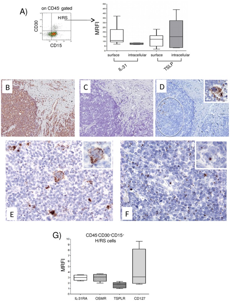
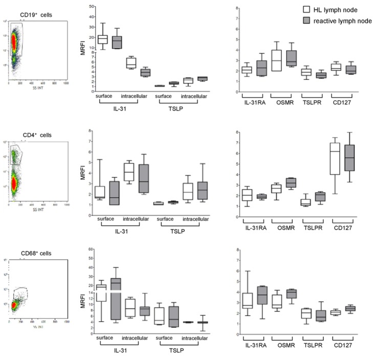
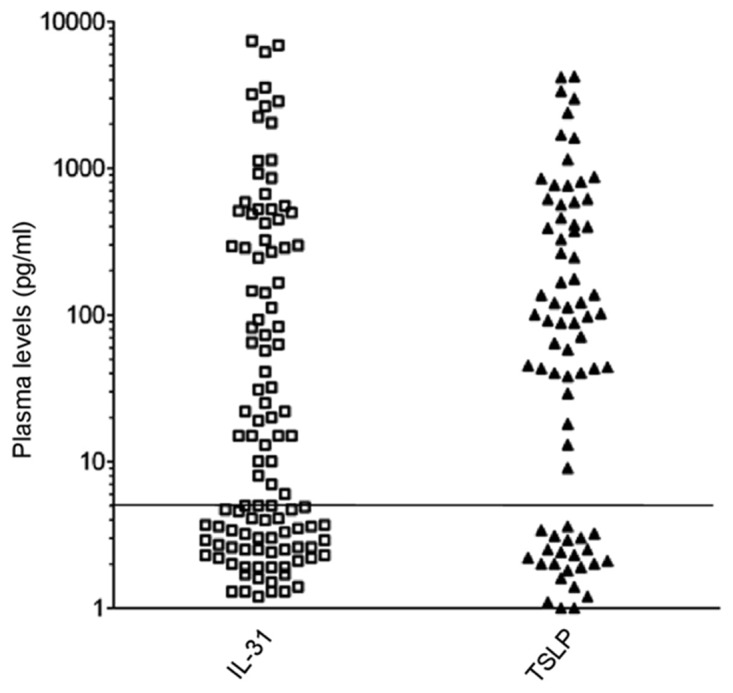
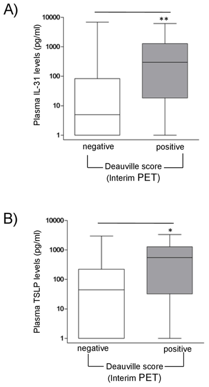
Hodgkin Lymphoma (HL) is a tumor of B-cell origin characterized by Hodgkin and Reed-Stenberg (H/RS) cells embedded in an inflammatory tissue where numerous cytokines/chemokines contribute to shape the microenvironment, leading to the typical clinical symptoms. We investigated: i) the expression of Interleukin-IL-31 (IL-31) and Thymic Stromal Lymphopoietin (TSLP), two Th2-related cytokines with tumor-promoting and pruritogenic functions, and of the respective receptors in HL invaded lymph nodes by flow cytometry, and ii) the potential association of IL-31/TSLP plasma concentrations with clinical characteristics by ELISA. H/RS cells and the major immune cell types infiltrating HL lymph nodes expressed intracytoplasmic and surface IL-31/TSLP, and their receptors. A subgroup of patients showing at diagnosis elevated IL-31 and TSLP plasma levels had an International Prognostic Score>2, indicative of high risk of relapse, and a subsequent positive interim PET-scan, indicative of insufficient response to chemotherapy. No correlation was found between IL-31/TSLP plasma levels and overall or event-free survival. In conclusion, IL-31/TSLP and their receptors are expressed in HL cells and in immune cells infiltrating affected lymph nodes, where both cytokines may contribute to local immune suppression. The clinical impact of IL-31 and TSLP plasma levels has to be further defined in larger patient cohorts.

Keywords: Hodgkin lymphoma; IL-31; PET; TSLP; cytokine receptors.


Conflict of interest statement

CONFLICTS OF INTEREST The authors declare that they have no competing financial interests in relation to the work described.

Figures

Figure 1
Figure 1. Expression of IL-31, TSLP and their receptors in H/RS cells
(A) Left panel. A representative gating strategy for H/RS cells identified as CD45, CD30+, CD15+ cells. Right panel. IL-31/TSLP expression was tested by flow cytometry at surface and intracellular levels. Results are expressed in box plot as median MRFI, first and third quartiles, maximum and minimum values, from 10 different HL lymph node cell suspensions. (B-F) In situ hybridization for Ubiquitin (B), dapB (C), CD30 (D), IL-31 (E) and TSLP (F) mRNA in cHL using the RNAscope technology (B, C, D) original magnification x100; E, F x200; insets x400). Ubiquitin mRNA was diffusely expressed (brown dots), whereas the bacterial dapB was completely negative. The CD30 probe hybridized with a proportion of the cells with H/RS morphology (circle and inset). Both IL-31 and TSLP mRNA were detected in the cytoplasm of H/RS cells (inset) and in some of the immune reactive cells present in the background. (G) IL-31RA/OSMR and TSLPR/CD127 chain receptor expression was analyzed by flow cytometry. Results are expressed in box plot as median MRFI, first and third quartiles, maximum and minimum values, from 7 different HL lymph node cell suspensions.
Figure 2
Figure 2. Expression of IL-31, TSLP and their receptors in lymphoid cells populating HL and reactive lymph node microenvironment
Left panels. A representative gating strategy for B cells (CD19+), T cells (CD4+), and macrophages (CD68+). Middle panels. IL-31/TSLP expression was analyzed by flow cytometry at surface and intracellular levels on CD19+, CD4+, and CD68+ cells for both HL and reactive lymph nodes. Results are shown in box plot as median MRFI, first and third quartiles, maximum and minimum values, from 7 different HL and 7 reactive lymph node cell suspensions. Right panels. IL-31RA/OSMR and TSLPR/CD127 chain receptor expression was analyzed by flow cytometry on CD19+, CD4+ and CD68+ cells for both HL and reactive lymph nodes. Results are shown in box plot as median MRFI, first and third quartiles, maximum and minimum values, from 7 different HL and 7 reactive lymph node cell suspensions.
Figure 3
Figure 3. sIL-31/TSLP plasma levels in HL patients
sIL-31/TSLP levels were assayed by ELISA (threshold of detection 5 pg/ml). sIL-31 was detected in 65/109 patients, sTSLP in 52/75 patients. The horizontal line separates positive from negative results.
Figure 4
Figure 4. Correlations between sIL-31/sTSLP and Interim Pet in HL patients
sIL-31 (A) and sTSLP (B) levels (pg/ml) in HL patient plasma were correlated with interim PET scan. PET images were classified according to the Deauville score (1-3 negative; 4-5 positive). HL patients with a positive score had significant higher median levels of sIL-31 and sTSLP than patients with negative score (**P=0.01, n=13/84 and *P=0.05, n=10/63, respectively).

References

    1. Campo E, Swerdlow SH, Harris NL, Pileri S, Stein H, Jaffe ES. The 2008 WHO classification of lymphoid neoplasms and beyond: evolving concepts and practical applications. Blood. 2011;117:5019–5032. - PMC - PubMed
    1. Aldinucci D, Gloghini A, Pinto A, De Filippi R, Carbone A. The classical Hodgkin's lymphoma microenvironment and its role in promoting tumour growth and immune escape. J Pathol. 2010;221:248–263. - PubMed
    1. Liu Y, Sattarzadeh A, Diepstra A, Visser L, van den Berg A. The microenvironment in classical Hodgkin lymphoma: an actively shaped and essential tumor component. Semin Cancer Biol. 2014;24:15–22. - PubMed
    1. Marafioti T, Hummel M, Foss HD, Laumen H, Korbjuhn P, Anagnostopoulos I, Lammert H, Demel G, Theil J, Wirth T, Stein H. Hodgkin and reed-sternberg cells represent an expansion of a single clone originating from a germinal center B-cell with functional immunoglobulin gene rearrangements but defective immunoglobulin transcription. Blood. 2000;95:1443–1450. - PubMed
    1. Re D, Kuppers R, Diehl V. Molecular pathogenesis of Hodgkin's lymphoma. J Clin Oncol. 2005;23:6379–6386. - PubMed

LinkOut - more resources