. 2017 Aug 3;8(49):85415-85427.
doi: 10.18632/oncotarget.19960. eCollection 2017 Oct 17.

A RasGAP, DAB2IP, regulates lipid droplet homeostasis by serving as GAP toward RAB40C

Affiliations

A RasGAP, DAB2IP, regulates lipid droplet homeostasis by serving as GAP toward RAB40C

Xiaomin Luo et al. Oncotarget. .

Erratum in

Abstract

Lipid droplet (LD) homeostasis involves activities of various RAB small GTPases. Recently, we found RAB40C was one of the RAB proteins regulating LD homeostasis. RAB40C contains a unique SOCS domain that is required for clustering of LDs. However, its precise functional role in LD homeostasis and mechanism of regulation remain largely unknown. In this study, we observed over-accumulation of LDs in cells with RAB40C deleted by Crispr-Cas9 editing. RAB40C appeared to reduce LD accumulation after long term incubation of cells with oleic acid (24 hours). Unexpectedly, we found that Ras GTPase activating protein (GAP), DAB2IP, bound to RAB40C mainly via its GAP domain and could serve as RAB40C GAP. Studies involving overexpression of DAB2IP and its GAP defective mutant and siRNA depletion of DAB2IP all confirmed that DAB2IP negatively regulated the effect of RAB40C on LD homeostasis. These results provide a novel perspective on the regulation of RAB40C and implicate various signalling pathways regulated by DAB2IP, which may play a role in LD homeostasis via RAB40C.

Keywords: DAB2IP; GTPase activating protein; RAB40C; lipid droplets.


Conflict of interest statement

CONFLICTS OF INTEREST The authors declare no competing financial interest.

Figures

Figure 1
Figure 1. RAB40C deletion caused over-accumulation of lipid droplet
The cells were fed with 40 μM oleic acid for 24 hours before fixation and staining. (A) Phase contrast images of wildtype HEK293T or various RAB40C mutant cell lines cultured in growth medium. (B) Bodipy493/503 staining in HEK293T cells of wildtype (Rab40+/+), RAB40C heterozygous (RAB40C+/m) and homozygous(RAB40Cm/m) mutant cell lines. Scale bar = 10μm. (C) Quantified average fluorescent signals of Bodipy493/503 per cell in each sample in panel B. N≥36 per sample. Error bar = SEM. ** p < 0.01, *** p<0.001. (D) Bodipy493/503 staining in hepatocytesLO2 with the indicated genotypes with respect to RAB40C. Scale bar = 10μm. (E) Quantified average fluorescent signals of Bodipy493/503 per cell in each sample in panel D. N≥40 per sample. Error bar = SEM, *** p<0.001.
Figure 2
Figure 2. RAB40C reduced LD accumulation in LO2 cells
(A) LO2 cells transfected with dsRed-RAB40C were incubated with oleic acid for the indicated time periods before fixed and staining with Bodipy493/503 for LDs. RAB40C transfected cells were marked with asterisks. Scale bar = 10 μm. (B) Quantification of the average Bodipy493/503 fluorescence signal per cell for the experiments shown in panel A. N ≥ 33. Error bars = SEM. *** p<0.001. (C) Bodipy493/503 staining of LO2 cells transfected with DsRed vector (top panels) or DsRed-RAB40C (bottom panels). (D) Quantification of the Bodipy493/503 fluorescence signal in DsRed vector or DsRed-RAB40C transfected and non-transfected cells. mDsRed-C1(-) indicates non-transfected cells in the sample transfected with DsRed vector. mDsRed-RAB40C(-) indicates non-transfected cells in the sample transfected with DsRed-RAB40C. N ≥ 30. Error bar = SEM. *** P< 0.001. In these experiments, the cells were fed with 400 μM OA for indicated time points in (A), and for 36 hours in (C).
Figure 3
Figure 3. Re-introduction of RAB40C rescued the LD over-accumulation in RAB40Cm/m cells in a nucleotide-dependent manner
(A) Bodipy493/503 staining in LO2 hepatocytes of the indicated genotypes transfected with mDsRed-RAB40C or mDsRed vector control. The cells were fed with 120 μM of oleic acid for 36 hours before fixation and staining. Transfected cells were indicated with asterisks. Scale bar = 10μm. (B) Quantified average fluorescent signals of Bodipy493/503 per cell in each sample in panel A. N≥30 per sample. Error bar = SEM. *** p<0.001. (C) Bodipy493/503 staining in RAB40Cm/m LO2 cells transfected with the wildtype DsRed-RAB40C, or GDP-locked (G28N), GTP-locked (Q73L), or SOCS mutant. Transfected cells were indicated with asterisks. Scale bar = 10 μm. (D) Quantified average fluorescent signals of Bodipy493/503 per cell in each sample. N ≥ 30 per sample. Error bar = SEM. *** p < 0.001.
Figure 4
Figure 4. RAB40C interacted with DAB2IP
(A) Myc-DAB2IP interacted with 3HA-RAB40C, but not with 3HA-RAB18. HEK293T cells transfected with the indicated expression plasmids were subjected to immunoprecipitation with anti-c-Myc (9E10) (left panels) or anti-HA (C5) (right panels), and the presence of co-purified proteins were detected by immunoblotting. (B) Lysates from HEK293T cells overexpessing Myc-DAB2IP were separately incubated with beads loaded with GST, GST-RAB40C and GST-RAB18. The presence of Myc-DAB2IP was detected by immunoblotting with anti-c-Myc antibody (left panel). Ponceau S staining of the same blot indicated the amount of GST proteins used for the pull down assay (right panel). (C) Immunoprecipitation of endogenous DAB2IP with anti-DAB2IP antibody also precipitated overexpressed RAB40C.
Figure 5
Figure 5. DAB2IP antagonizes RAB40C reduction of LD accumulation
(A) Bodipy493/503 staining in hepatocytes LO2 overexpressed with mDsRed-RAB40C (top panel), Myc-DAB2IP (second panel), both RAB40C and DAB2IP (third panel), GAP defective DAB2IP mutant R385L (fourth panel), and co-expression with R385L and RAB40C (bottom panel). Cells expressing the indicated Myc-tagged fusion proteins are labeled with asterisks. Cells co-expressing both indicated proteins are labeled with empty triangles. The cells were fed with 400 μM of oleic acid for 36 hours before fixation and staining Scale bar = 10 um. (B) Quantification of Bodipy493/503 staining of samples shown in A. N ≥ 30. ** p < 0.01; *** p < 0.001. (C) siRNA depletion of DAB2IP in LO2 hepatocytes. Lysates from LO2 cells non-transfected (uT), or transfected with negative control siRNA (NCsi), or with siRNA duplexes specific to DAB2IP (DAB2IPsi were subjected to immunoblotting to determine the efficiency of DAB2IP knockdown. GAPDH serves as a loading control (bottom panel). (D) Bodipy493/503 staining in LO2 hepatocytes silenced with control siRNA (left two panels), or siRNA DAB2IPsi-1 (middle two), or DAB2IPsi-1 co-expressed with DsRed-RAB40C (right three). (E) Quantification of the Bodipy493/503 stainings of the samples shown in panel D. N ≥ 30. Error bar = SEM. ***P< 0.001.
Figure 6
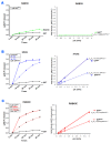
Figure 6. DAB2IP stimulated GTP hydrolysis of RAB40C
(A) GAP domain of DAB2IP did not stimulate GTP hydrolysis of Rab18. GTP hydrolysis by GST-Rab18 (green line) was measured by the release of Pi as a function of time (left panel). GST (black line) was used as negative control. Catalytic efficiencies of DAB2IP toward Rab18 and GST were analyzed by Kcat/Km plot (right panel). (B) DAB2IP GAP domain stimulated GTP hydrolysis of H-Ras. GTP hydrolysis by GST-H-Ras was stimulated by wildtype GAP domain (solid blue line) or GAP defective mutant R385L (dotted blue line) as a function of time (left panel). Catalytic efficiencies of wildtype (solid blue line) and R385L (dotted blue line) GAP domains toward H-Ras were analyzed by Kcat/KM plot (right panel). (C) DAB2IP GAP domain stimulated GTP hydrolysis of RAB40C. GTP hydrolysis by GST-RAB40C was stimulated by wildtype GAP domain (solid blue line) or GAP defective mutant R385L (dotted blue line) as a function of time (left panel). Catalytic efficiencies of wildtype (solid blue line) and R385L (dashed blue line) GAP domains toward RAB40C were analyzed by Kcat/KM plot (right panel). All data points were averages of three independent experiments. Error bars = SEM.

References

    1. Wang B, Li L, Fu J, Yu P, Gong D, Zeng C, Zeng Z. Effects of long-chain and medium-chain fatty acids on apoptosis and oxidative stress in human liver cells with steatosis. J Food Sci. 2016;81:H794–800. https://doi.org/10.1111/1750-3841.13210. - DOI - PubMed
    1. Khan SA, Wollaston-Hayden EE, Markowski TW, Higgins L, Mashek DG. Quantitative analysis of the murine lipid droplet-associated proteome during diet-induced hepatic steatosis. J Lipid Res. 2015;56:2260–72. https://doi.org/10.1194/jlr.M056812. - DOI - PMC - PubMed
    1. Wang W, Wei S, Li L, Su X, Du C, Li F, Geng B, Liu P, Xu G. Proteomic analysis of murine testes lipid droplets. Sci Rep. 2015;5:12070. https://doi.org/10.1038/srep12070. - DOI - PMC - PubMed
    1. O’Mahony F, Wroblewski K, O’Byrne SM, Jiang H, Clerkin K, Benhammou J, Blaner WS, Beaven SW. Liver X receptors balance lipid stores in hepatic stellate cells through Rab18, a retinoid responsive lipid droplet protein. Hepatology. 2015;62:615–26. https://doi.org/10.1002/hep.27645. - DOI - PMC - PubMed
    1. Tang WC, Lin RJ, Liao CL, Lin YL. Rab18 facilitates dengue virus infection by targeting fatty acid synthase to sites of viral replication. J Virol. 2014;88:6793–804. https://doi.org/10.1128/jvi.00045-14. - DOI - PMC - PubMed