. 2017 Aug 14;8(49):85537-85548.
doi: 10.18632/oncotarget.20246. eCollection 2017 Oct 17.

Electroacupuncture alleviates neuromuscular dysfunction in an experimental rat model of immobilization

Affiliations

Electroacupuncture alleviates neuromuscular dysfunction in an experimental rat model of immobilization

Jun Yang et al. Oncotarget. .

Abstract

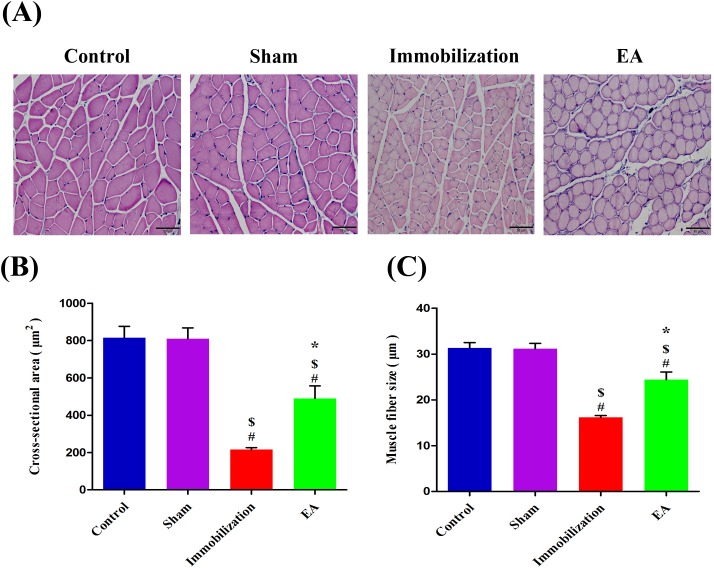
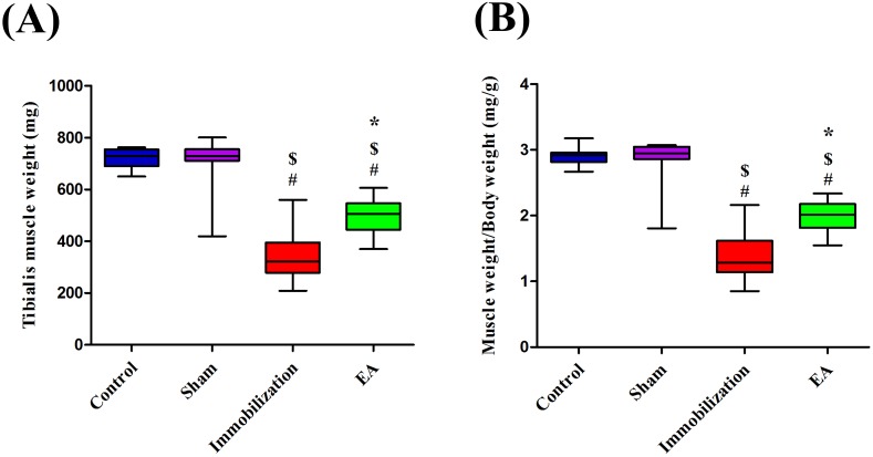
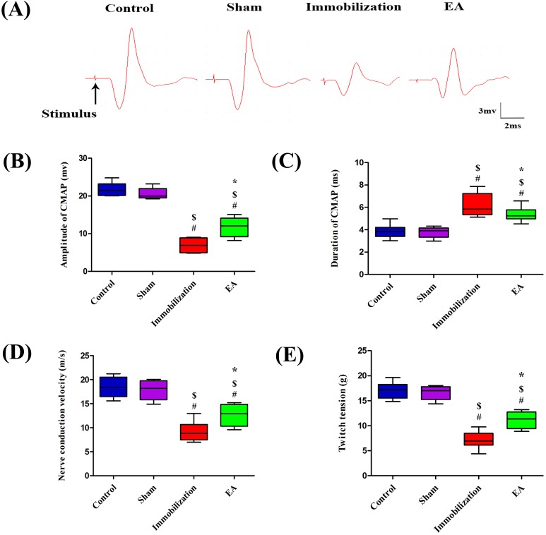
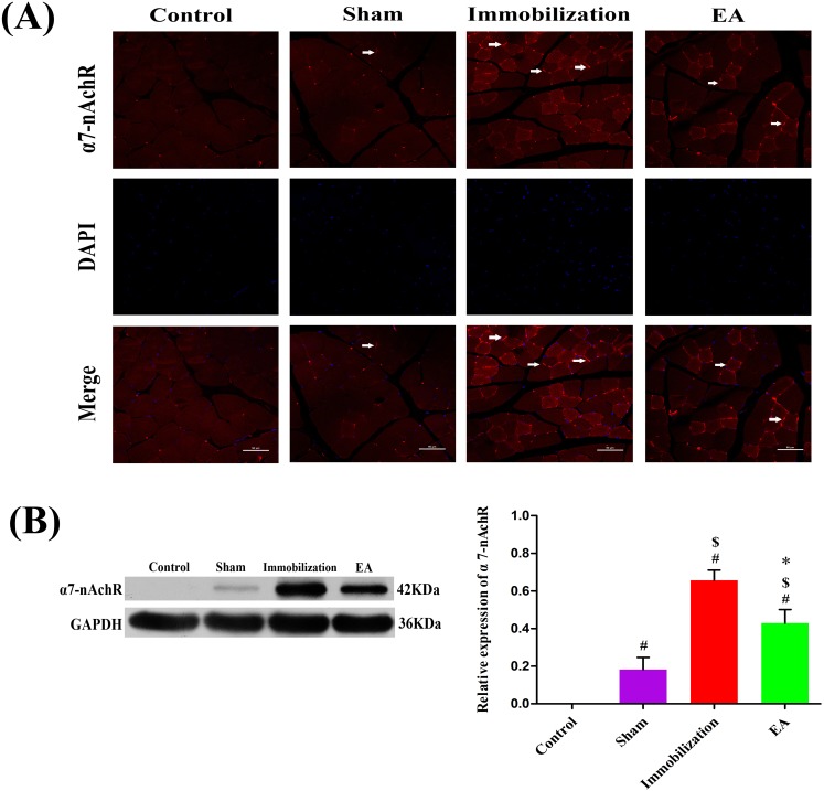
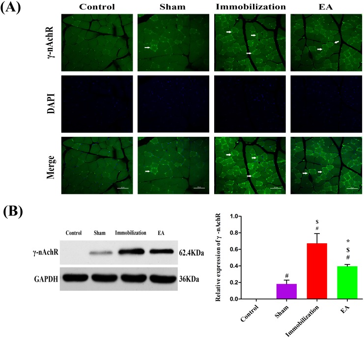
Immobilization-related skeletal muscle atrophy is a major concern to patients in Intensive Care Units and it has a profound effect on the quality of life. However, the underlying molecular events for the therapeutic effect of electroacupuncture to treat muscle atrophy have not been fully elucidated. Here we developed an immobilization mouse model and tested the hypothesis that skeletal muscle weakness may be caused by the increased expression of γ and α7 nicotinic acetylcholine receptors (nAChRs) on muscle cell membranes, while electroacupuncture could decrease the expression of γ and α7 nicotinic acetylcholine receptors. Compared with the rats in control, those treated with immobilization for 14 days showed a significant reduction of tibialis anterior muscle weight, muscle atrophy and dysfunction, which was associated with a significant decrease expression of neuregulin-1 and increased expression of γ- and α7-nAChR in tibialis anterior muscle. Electroacupuncture significantly enhanced the expression of neuregulin-1 and alleviated the muscle loss, while diminished the expression of γ- and α7-nAChR. Taken together, the beneficial effect of electroacupuncture may be attributed to suppressing γ- and α7-nAChR production, enhancing neuromuscular function and neuregulin-1 protein synthesis. These results suggest that electroacupuncture is a potential therapy for preventing muscle atrophy during immobilization.

Keywords: electroacupuncture; immobilization; neuromuscular function; nicotinic acetylcholine receptors; skeletal muscle atrophy.


Conflict of interest statement

CONFLICTS OF INTEREST The authors declare no competing financial interests.

Figures

Figure 1
Figure 1. EA prevents immobilization-induced muscle fiber cross-sectional area and muscle fiber size decrease
(A) The morphological changes of tibialis anterior muscle stained with H&E (magnification, ×200, scale bar = 50μm) in four groups. (B, C) After 14 days immobilization, muscle fiber Cross-sectional area andmuscle fiber size were significantly decreased in the Immobilization group than in the Control group (P < 0.05). With EA treatment, Cross-sectional area andmuscle fiber size were significantly greater in the EA group than in the Immobilization group (P < 0.05). The average size of myofibers determined from six mice x 10 sections/mouse/group (Bars: means ± SD; n = 6/group). P < 0.05 is significant (# vs. Control, $ vs. Sham, *vs. Immobilization).
Figure 2
Figure 2. EA prevents immobilization-induced muscle atrophy
(A) Box plot shows Tibialis anterior muscle weight. (B) Box plot shows muscle wet weight/body weight ratio at different groups. (n=8/group). # P < 0.05 vs. Control group; $ P < 0.05 vs. Sham group; * P < 0.05 vs. Immobilization group.
Figure 3
Figure 3. EA improves neuromuscular function
CMAP recorded in the tibialis anterior muscle by needle electrodes after supramaximal needle electrode stimulation of the sciatic nerve. (A) Samples of CMAP recorded in each group, (B) amplitudes, (C) durations, (D) NCV, and (E) single-twitch tension elicited by continuous indirect stimulation. (n=8/group). # P < 0.05 vs. Control group; $ P < 0.05 vs. Sham group; * P < 0.05 vs. Immobilization group.
Figure 4
Figure 4. HCA and PCA analysis of neuromuscular function
(A) HCA, Hierarchical cluster analysis of the neuromuscular function of rats. The neuromuscular function of rats in the Control, Sham, Immobilization and the EA group was labeled as C1∼C8, S1∼S8, Im1∼Im8 and EA1∼EA8. (B) PCA scores of neuromuscular function of rats. (▲, Control group; ▼, Sham group; ●, Immobilization group; ★, EA group; n=8/group).
Figure 5
Figure 5. Effects of EA on the expression of α7-nAChR
(A) Immunofluorescence analysis was performed to determine α7-nAChR expression on Tiabilis anterior muscle 14 days after the procedure. The samples were immunostained with an anti-α7-nAChR-antibody (red, → marks a positive expression). The result shows that α7-nAChR expression are sharply increased and clustered on the muscle membrane of rats in the immobilization group. Representative results from three independent experiments are shown here (scale bar = 50 μm). (B) The Western blot analysis of the α7-nAChR proteins are shown for each groups. Relative intensity of α7-nAChR to GAPDH is shown in the graphs. α7-nAChR significantly increased after the immobilization for 14 days. Electroacupuncture suppressed the expression of α7-nAChR in EA group. All values are expressed as means ± SD (n=6/group).# P < 0.05 vs. Control group; $ P < 0.05 vs. Sham group; * P < 0.05 vs. Immobilization group.
Figure 6
Figure 6. Effects of EA on the expression of γ-nAChR
(A) Immunofluorescence analysis was performed to determine γ-nAChR expression on Tiabilis anterior muscle 14 days after the procedure. The samples were immunostained with an anti-γ-nAChR-antibody (green, → marks a positive expression). The result shows that γ-nAChR expression are sharply increased and clustered on the muscle membrane of rats in the immobilization group. Representative results from three independent experiments are shown here (scale bar = 50μm). (B) The Western blot analysis of the γ-nAChR proteins are shown for each groups. Relative intensity of γ-nAChR to GAPDH is shown in the graphs.γ-nAChR significantly increased after the immobilization for 14 days. Electroacupuncture inhibited the expression of γ-nAChR in EA group. All values are expressed as means ± SD (n=6/group).# P < 0.05 vs. Control group; $ P < 0.05 vs. Sham group; * P < 0.05 vs. Immobilization group.
Figure 7
Figure 7. EA activates NRG-1 synthesis in the tiabilis anterior muscle of immobilization rats
Western blot analysis indicated that NRG-1 protein levels in the disuse muscles significantly decreased after 14 days of immobilization compared with the control group (P < 0.05). Densitometric analysis showed a significant increase in NRG-1 in the tiabilis anterior muscle after electroacupuncture therapy compared with the immobilization group (P < 0.05). The integrated optical density of NRG-1 normalized to integrated optical density of GAPDH. The data are shown as means ± SD (n=6/group).# P < 0.05 vs. Control group; $ P < 0.05 vs. Sham group; * P < 0.05 vs. Immobilization group.

Similar articles

Cited by

References

    1. Walsh CJ, Batt J, Herridge MS, Mathur S, Bader GD, Hu PZ, dos Santos CC. Transcriptomic analysis reveals abnormal muscle repair and remodeling in survivors of critical illness with sustained weakness. Scientific reports. 2016;6:29334. - PMC - PubMed
    1. Truong AD, Fan E, Brower RG, Needham DM. Bench-to-bedside review: mobilizing patients in the intensive care unit from pathophysiology to clinical trials. Critical Care. 2009;13:4–216. - PMC - PubMed
    1. Cohen S, Nathan JA, Goldberg AL. Muscle wasting in disease: molecular mechanisms and promising therapies. Nat Rev Drug Discov. 2015;14:58–74. - PubMed
    1. McNally EM, Pytel P. Muscle diseases: the muscular dystrophies. Annual review of pathology. 2007;2:87–109. - PubMed
    1. Robinson GA, Enoka RM, Stuart DG. Immobilization-induced changes in motor unit force and fatigability in the cat. Muscle & nerve. 1991;14:563–573. - PubMed

LinkOut - more resources