Review
. 2018 Feb;28(1):55-65.
doi: 10.1016/j.nic.2017.09.002. Epub 2017 Oct 23.

Perfusion Imaging in Acute Traumatic Brain Injury

Affiliations
Review

Perfusion Imaging in Acute Traumatic Brain Injury

David B Douglas et al. Neuroimaging Clin N Am. 2018 Feb.

Abstract

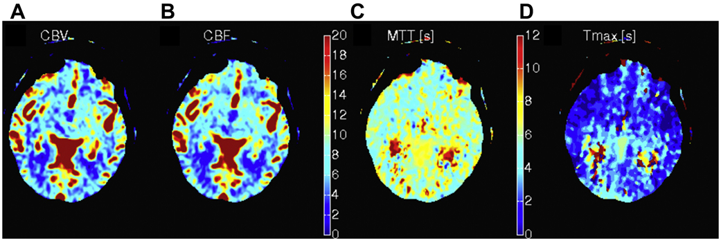
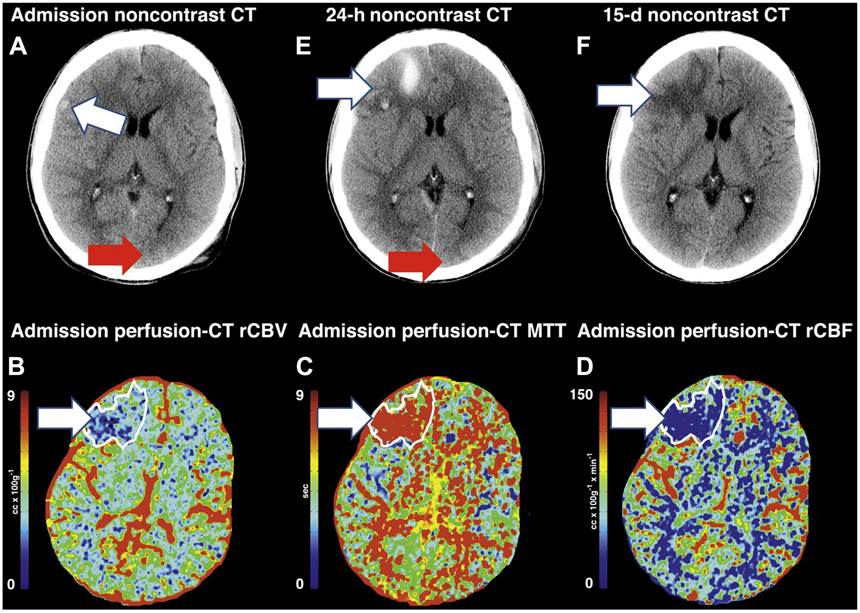
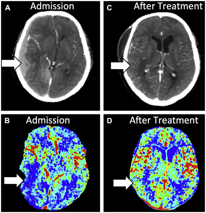
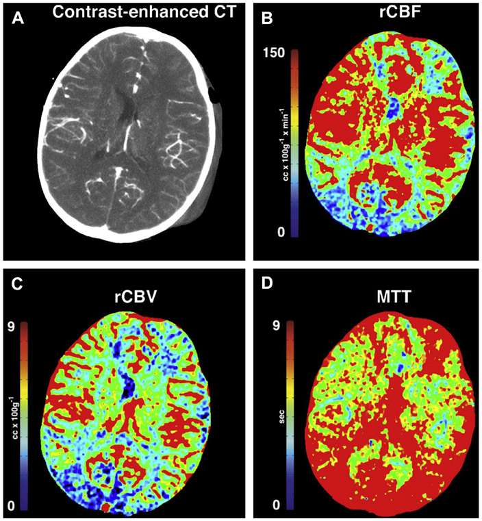
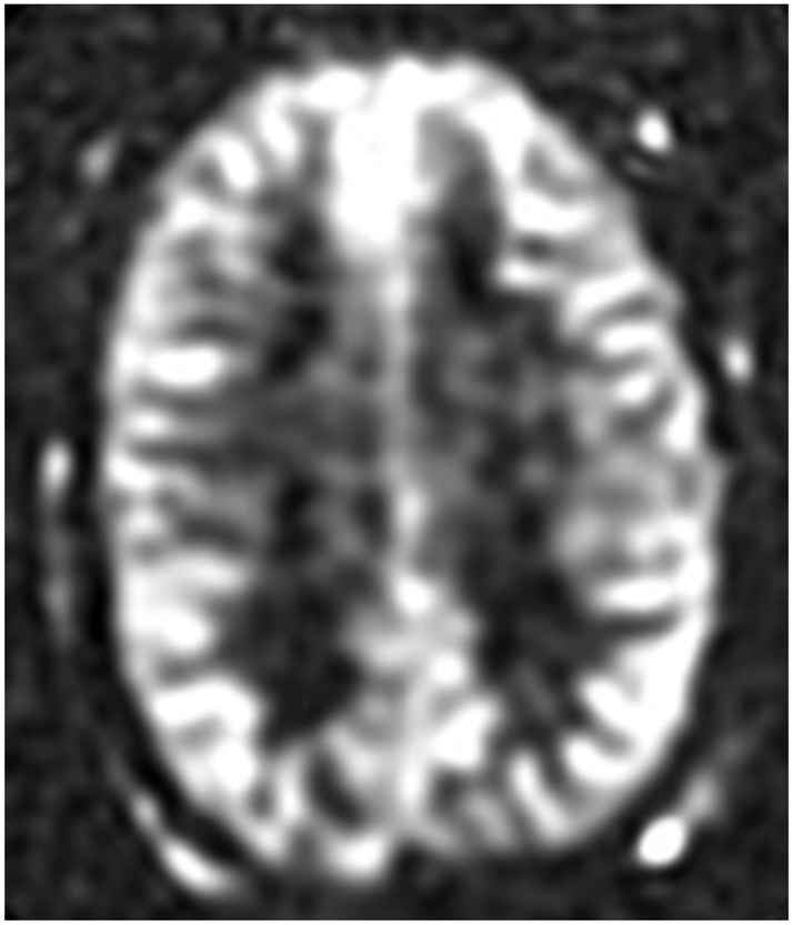
Traumatic brain injury (TBI) is a significant problem worldwide and neuroimaging plays a critical role in diagnosis and management. Recently, perfusion neuroimaging techniques have been explored in TBI to determine and characterize potential perfusion neuroimaging biomarkers to aid in diagnosis, treatment, and prognosis. In this article, computed tomography (CT) bolus perfusion, MR imaging bolus perfusion, MR imaging arterial spin labeling perfusion, and xenon CT are reviewed with a focus on their applications in acute TBI. Future research directions are also discussed.

Keywords: Concussion; Perfusion; TBI; Traumatic brain injury.


Figures

Figure 1:
Figure 1:
Normal CT Perfusion images showing (A) rCBV, (B) rCBF, (C) MTT and (D) Tmax.
Figure 2:
Figure 2:
Contrast-enhanced and perfusion-CT images from a patient with severe TBI. (A) Noncontrast CT at admission demonstrates a small hemorrhagic contusion in the right frontal lobe (arrow). Admission perfusion-CT images demonstrate a large territory of decreased rCBV (B), increased MTT (C) and decreased rCBF (D). Follow-up noncontrast CT at 24 hours (E) demonstrates increased areas of hemorrhagic contusion in the right frontal lobe where the perfusion abnormality was seen. Follow-up noncontrast CT at 15-days (F) demonstrates evolving hemorrhagic contusion and encephalomalacia in the right frontal lobe, which corresponds to the same distribution that is seen on the perfusion-CT.
Figure 3:
Figure 3:
Contrast-enhanced and PCT images from a patient with severe TBI. (A) Contrast-enhanced CT imaging at admission demonstrates a right-sided subdural hematoma causing mass effect on the underlying brain and midline shift. (B) rCBF PCT imaging at admission demonstrates decreased rCBF in the right temporal lobe. (C) Contrast-enhanced CT image after surgical evacuation of the hematoma demonstrates resolution of the right-sided subdural hematoma, mass effect and midline shift. (D) rCBV PCT imaging after surgical evacuation of the right-sided hematoma demonstrates normalization of the rCBF in the right temporal lobe.
Figure 4:
Figure 4:
Contrast-enhanced and PCT images of a case of TBI with intracranial hypertension. The contrast enhanced CT (A) demonstrated left-sided scalp hematoma. The rCBF (B) and rCBV (C) trended toward lower values especially in the occipital lobes. The MTT (D) demonstrated significantly higher values, reflecting altered cerebral autoregulation after TBI.
Figure 5:
Figure 5:
Normal noncontrast arterial spin labeling (ASL) sequence.
Figure 6:
Figure 6:
Two Tc-99m-HMPAO images of a Molecular Neuroimaging of Cerebral Blood Flow Abnormalities due to Traumatic Brain Injury (TBI) in a Swine Model (Sus acrofa). Control (A) illustrates normal perfusion (red arrow). The 1-hour post-TBI (B) illustrates craniectomy site (white arrow) at the top of the image with hypoperfusion seen in the underlying brain (red arrow).

References

    1. Faul M, National Center for Injury Prevention and Control (U.S.). Traumatic brain injury in the United States : emergency department visits, hospitalizations, and deaths, 2002-2006. http://purl.fdlp.gov/GPO/gpo41911.
    1. Marin JR, Weaver MD, Yealy DM, Mannix RC. Trends in visits for traumatic brain injury to emergency departments in the United States. Jama. 2014;311(18):1917–1919. - PubMed
    1. Centers for Disease C, Prevention. Nonfatal traumatic brain injuries related to sports and recreation activities among persons aged </=19 years--United States, 2001-2009. MMWR Morbidity and mortality weekly report. 2011;60(39):1337–1342. - PubMed
    1. Corrigan JD, Selassie AW, Orman JA. The epidemiology of traumatic brain injury. The Journal of head trauma rehabilitation. 2010;25(2):72–80. - PubMed
    1. Bass E, Golding H, United States. Congressional Budget Office. The Veterans Health Administration's treatment of PTSD and traumatic brain injury among recent combat veterans. A CBO study. Washington, DC: Congress of the United States, Congressional Budget Office,; 2012: http://purl.fdlp.gov/GPO/gpo18872.