Review
. 2018 Jan 5;373(1737):20160479.
doi: 10.1098/rstb.2016.0479.

Why the need and how to approach the functional diversity of extracellular vesicles

Affiliations
Review

Why the need and how to approach the functional diversity of extracellular vesicles

Mercedes Tkach et al. Philos Trans R Soc Lond B Biol Sci. .

Abstract

In the past decade, cell-to-cell communication mediated by exosomes has attracted growing attention from biomedical scientists and physicians, leading to several recent publications in top-tier journals. Exosomes are generally defined as secreted membrane vesicles, or extracellular vesicles (EVs), corresponding to the intraluminal vesicles of late endosomal compartments, which are secreted upon fusion of multi-vesicular endosomes with the cell's plasma membrane. Cells, however, were shown to release other types of EVs, for instance, by direct budding off their plasma membrane. Some of these EVs share with exosomes major biophysical and biochemical characteristics, such as size, density and membrane orientation, which impose difficulties in their efficient separation. Despite frequent claims in the literature, whether exosomes really display more important patho/physiological functions, or are endowed with higher potential as diagnostic or therapeutic tools than other EVs, is not yet convincingly demonstrated. In this opinion article, we describe reasons for this lack of precision knowledge in the current stage of the EV field, we review recently described approaches to overcome these caveats, and we propose ways to improve our knowledge on the respective functions of distinct EVs, which will be crucial for future development of well-designed EV-based clinical applications.This article is part of the discussion meeting issue 'Extracellular vesicles and the tumour microenvironment'.

Keywords: cancer; exosomes; extracellular vesicles; multi-vesicular endosomes.


Conflict of interest statement

We declare we have no competing interests.

Figures
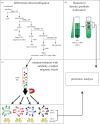
Figure 1.
Figure 1.
Different approaches to analyse the heterogeneity of EVs (based on [26]). (a) Scheme of EV isolation by dUC protocol from a mixture of EVs present in cell culture conditioned medium or body fluids. For the latter EV source, additional steps of biofluid processing before centrifugation are recommended to eliminate abundant EV-excluded components. (b) Further separation of the 10 K and 100 K pellets obtained by the dUC protocol through flotation in a density gradient (iodixanol) allowed us to distinguish discrete populations of EVs by proteomic analysis. (c) Separation of subtypes of small EVs through immunoisolation. We isolated small EVs bound specifically to beads coupled to antibodies to CD9, or to CD63, or to CD81 or to irrelevant IgG as control. We analysed these EV subpopulations by proteomic analysis in parallel with the non-pulled down materials remaining in the flow-through for each immunoisolation. Comparing EVs pulled down via the different tetraspanin-specific antibodies identified an exosomal EV subpopulation as bearing CD63 together with the other tetraspanins. Analysing the flow-through demonstrated the presence of non-exosomal small EVs in the 100 K pellet.
Figure 2.
Figure 2.
Functional analysis of heterogeneous EVs populations. When analysing the functionality of EVs released by a cell line, a primary culture or even from body fluids, the results can be extremely biased depending on the isolation technique used. For example, in the scheme, we illustrate a case where both large blue EVs and small green EVs have a positive effect on the analysed target cells, while the large orange EVs and small yellow EVs have a negative effect. Depending of the ratio of these EVs in a given preparation, we can either observe a positive or a negative effect, or even no effect if the mixture of all EV subtypes compensates the respective effects.

References

    1. Wolf P. 1967. The nature and significance of platelet products in human plasma. Br. J. Haematol. 13, 269–288. (10.1111/j.1365-2141.1967.tb08741.x) - DOI - PubMed
    1. Anderson HC. 1969. Vesicles associated with calcification in the matrix of epiphyseal cartilage. J. Cell Biol. 41, 59–72. (10.1083/jcb.41.1.59) - DOI - PMC - PubMed
    1. Crawford N. 1971. The presence of contractile proteins in platelet microparticles isolated from human and animal platelet-free plasma. Br. J. Haematol. 21, 53–69. (10.1111/j.1365-2141.1971.tb03416.x) - DOI - PubMed
    1. Dalton AJ. 1975. Microvesicles and vesicles of multivesicular bodies versus ‘virus-like’ particles. J. Natl Cancer Inst. 54, 1137–1148. (10.1093/jnci/54.5.1137) - DOI - PubMed
    1. Trams EG, Lauter CJ, Salem N Jr, Heine U. 1981. Exfoliation of membrane ecto-enzymes in the form of micro-vesicles. Biochim. Biophys. Acta 645, 63–70. (10.1016/0005-2736(81)90512-5) - DOI - PubMed

MeSH terms