. 2017 Dec 5;114(49):13012-13017.
doi: 10.1073/pnas.1717506114. Epub 2017 Nov 20.

Retrograde inhibition by a specific subset of interpeduncular α5 nicotinic neurons regulates nicotine preference

Affiliations

Retrograde inhibition by a specific subset of interpeduncular α5 nicotinic neurons regulates nicotine preference

Jessica L Ables et al. Proc Natl Acad Sci U S A. .

Abstract

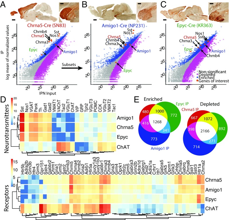
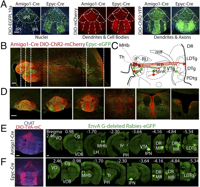
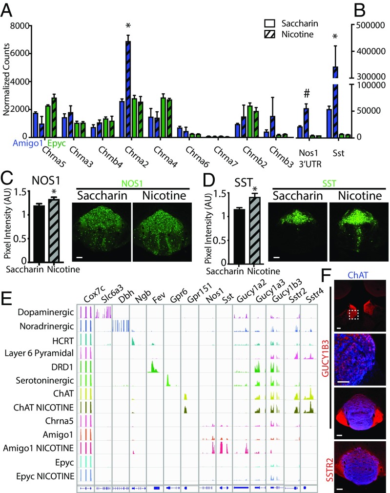
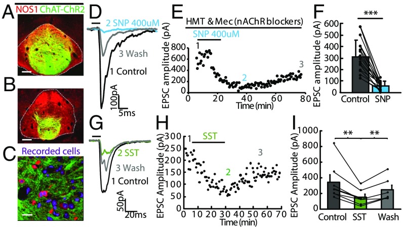
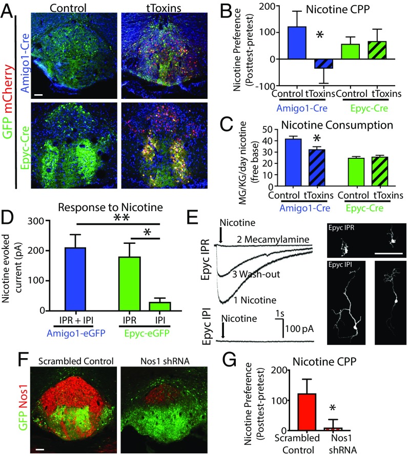
Repeated exposure to drugs of abuse can produce adaptive changes that lead to the establishment of dependence. It has been shown that allelic variation in the α5 nicotinic acetylcholine receptor (nAChR) gene CHRNA5 is associated with higher risk of tobacco dependence. In the brain, α5-containing nAChRs are expressed at very high levels in the interpeduncular nucleus (IPN). Here we identified two nonoverlapping α5 + cell populations (α5- Amigo1 and α5- Epyc ) in mouse IPN that respond differentially to nicotine. Chronic nicotine treatment altered the translational profile of more than 1,000 genes in α5- Amigo1 neurons, including neuronal nitric oxide synthase (Nos1) and somatostatin (Sst). In contrast, expression of few genes was altered in the α5- Epyc population. We show that both nitric oxide and SST suppress optically evoked neurotransmitter release from the terminals of habenular (Hb) neurons in IPN. Moreover, in vivo silencing of neurotransmitter release from the α5- Amigo1 but not from the α5- Epyc population eliminates nicotine reward, measured using place preference. This loss of nicotine reward was mimicked by shRNA-mediated knockdown of Nos1 in the IPN. These findings reveal a proaddiction adaptive response to chronic nicotine in which nitric oxide and SST are released by a specific α5+ neuronal population to provide retrograde inhibition of the Hb-IPN circuit and thereby enhance the motivational properties of nicotine.

Keywords: interpeduncular nucleus; nicotine; retrograde; α5 nicotinic.


Conflict of interest statement

The authors declare no conflict of interest.

Figures

Fig. 1.
Fig. 1.
TRAP profiling reveals two subsets of α5 neuronal populations in the IPN. (A) Chrna5-Cre mice (Top) crossed to EGFP-L10a reporter mice were used for TRAP. (Bottom) Scatterplots of average TRAP IP samples (y axis) versus input samples (x axis) represent enriched (>0.6 log2 fold change, blue) or depleted (less than −0.6 log2 fold change, magenta) translated mRNAs. Highly enriched mRNAs for Amigo1 and Epyc were identified for further characterization. (B) Amigo1-Cre::EGFP-L10a mice (C) and Epyc-Cre::EGFP-L10a mice were employed for TRAP and showed enriched mRNAs for Chrna5-Chrna3-Chrnb4 as indicated in the corresponding scatterplots. (AC) (Scale bar: low magnification 1 mm, high magnification 200 µm.) Images courtesy of GENSAT.org. (D) Expression levels (z-score transformed normalized counts) of receptors and neurotransmitters in the three IPN populations as well as in ChAT MHb neurons. (E) α5-Amigo1 and α5-Epyc cells share a large number of enriched and depleted mRNAs with Chrna5 cells.
Fig. 2.
Fig. 2.
α5-Amigo1 and α5-Epyc cells are two distinct, complementary IPN populations. (A) Amigo1-Cre and Epyc-Cre mice were injected with the indicated AAVs to label distinct cell compartments and determine the population distribution. α5-Amigo1 and α5-Epyc cells are present in the rostral subnucleus (IPR), but α5-Amigo1 cells are largely excluded from the intermediate subnuclei (IPI), where α5-Epyc cells are densely distributed. (Scale bar: 100 µm.) (B) Anterograde tracing in Epyc-EGFP × Amigo1-Cre mice injected with DIO-ChR2-mCherry shows dense Amigo1 projections (red) from IPN to caudal structures relative to Epyc projections (green). (Scale bar: 200 µm.) (C) Schematic representation of Amigo projections (red) and Epyc projections (green). Epyc cells appear to be a series of local interneurons along the IPN–raphe axis, with a relatively minor projection from IPN to raphe and caudal structures. (D) Coronal sections corresponding to the dashed lines in A. (Scale bar: 200 µm.) (E and F) Retrograde tracing with rabies virus in Amigo1-Cre and Epyc-Cre mice. Coronal sections of IPN from Amigo1-Cre (E) or Epyc-Cre (F) mouse injected with DIO-TVA-mCherry and DIO-rabies G protein (1:1, red, Left) followed by injection of EnvA G-deleted rabies-EGFP to label monosynaptic inputs (green, Right). (Scale bar: low magnification 500 µm, high magnification 100 µm.)
Fig. 3.
Fig. 3.
Chronic nicotine up-regulates SST and NOS1 in α5-Amigo1 cells. (A) Normalized RNA-Seq counts of nAChRs in Amigo1-Cre::EGFP-L10a (blue) and Epyc-Cre::EGFP-L10a (green) mice after 6 wk of saccharin (solid bars) or nicotine (striped bars). Note that only Chrna2 is up-regulated in α5-Amigo1 cells. (B) Normalized RNA-Seq counts of Nos1 3′ UTR and Sst in Amigo1 and Epyc mice after 6 wk of nicotine (*Padj < 0.05, #Padj = 0.08, DESeq2). (C) Quantification of IHC pixel intensity in the IPN confirms increased expression of NOS1 (C) and SST (D) after chronic nicotine (*P < 0.05, unpaired two-tailed t test). (Scale bar: 100 µm.) (E) Integrated Genomics Viewer (IGV 2.3) of cell-specific translated mRNAs. Top row indicates Cox7c, a housekeeping gene, followed by positive control genes expressed in each cell type, followed by Nos1 and Sst and their respective receptors Gucy1a2, Gucy1a3, Gucy1b3, Sstr2, and Sstr4. Note the specific increase in expression of Nos1 and Sst in Amigo1 mice after 6 wk of nicotine treatment. (F) GUCY1B3 is expressed in the cell bodies and terminals of cholinergic MHb cells, and SSTR2 in MHb terminals in the IPN. (Scale bar: low magnification 100 µm, high magnification 50 µm.)
Fig. 4.
Fig. 4.
NO and SST inhibit evoked neurotransmitter release from axonal terminals of MHb neurons to the IPN. (A and B) Coronal sections from a ChAT-ChR2-EYFP mouse showing expression of NOS1 (red) in ChR2-EYFP+ terminals (green) in the IPN. (Scale bar: 100 µm.) (C) High-magnification view shows that ChR2-EYFP–labeled axonal terminals (green) encircle NOS1 immunopositive neurons (red) in the IPN. (Scale bar: 20 µm.) (D and E) Representative electrophysiological traces (D) and plot of EPSC amplitude versus time (E) show that application of the NO donor sodium nitroprusside (SNP) (400 µM) into the bath solution completely abolishes fast EPSCs evoked by brief blue light illumination (5 ms; horizontal bar) in the presence of nAChR blockers hexamethonium (HMT) and mecamylamine (Mec) and this blockade lasted for over 1 h. (F) Summary data show that SNP eliminates EPSCs (***P < 0.001, paired t test, n = 15 cells). Separate lines represent data from individual neurons. Control indicates EPSCs measured in nAChRs blockers. (GI) Representative electrophysiological traces (G) and plot of EPSC amplitude versus time (H) show that application of SST (1 µM) reduces fast EPSCs evoked by brief blue light illumination (5 ms; horizontal bar) and this inhibition lasted for over 20 min in wash solution. (I) Summary data show that SST reduces EPSCs (**P < 0.005, paired t test, n = 6 cells). Separate lines represent data from individual neurons. Error bars indicate SEM.
Fig. 5.
Fig. 5.
Blocking neurotransmission of α5-Amigo1 cells or knocking down Nos1 in IPN prevents nicotine preference. (A) Viral expression in control and tToxin-injected mice. (Scale bar: 100 µm.) (B) Place preference for a rewarding dose of nicotine (0.35 mg/kg, free base, i.p.) was eliminated in Amigo1-Cre mice expressing tToxins but not Epyc-Cre mice (*P < 0.05, unpaired one-tailed t test). (C) Average daily nicotine consumption was reduced in Amigo1-Cre but not Epyc-Cre mice injected with tToxins compared with control (*P < 0.05, unpaired two-tailed t test). (D) Average current amplitude of nicotine-evoked responses in Amigo1 and Epyc cells located in IPR and IPI regions (*P < 0.05, **P < 0.01, unpaired two-tailed t test, n = 11, 7, 8 cells, respectively). (E) Representative nicotine currents in IPR and IPI Epyc neurons and photomicrographs of streptavidin-filled neurons. (Scale bar: 100 µm.) (F) NOS1 expression is reduced by >75% in WT animals injected with Nos1 shRNA but not with a scrambled control virus. (Scale bar: 100 µm.) (G) Knockdown of Nos1 in the IPN of WT mice blocked place preference for nicotine (0.35 mg/kg, free base, i.p., *P < 0.05, unpaired one-tailed t test).

References

    1. Klemm WR. Habenular and interpeduncularis nuclei: Shared components in multiple-function networks. Med Sci Monit. 2004;10:RA261–RA273. - PubMed
    1. Boulos LJ, Darcq E, Kieffer BL. Translating the habenula: From rodents to humans. Biol Psychiatry. 2017;81:296–305. - PMC - PubMed
    1. Antolin-Fontes B, Ables JL, Gorlich A, Ibanez-Tallon I. The habenulo-interpeduncular pathway in nicotine aversion and withdrawal. Neuropharmacology. 2015;96:213–222. - PMC - PubMed
    1. Bierut LJ, et al. Variants in nicotinic receptors and risk for nicotine dependence. Am J Psychiatry. 2008;165:1163–1171. - PMC - PubMed
    1. Thorgeirsson TE, et al. A variant associated with nicotine dependence, lung cancer and peripheral arterial disease. Nature. 2008;452:638–642. - PMC - PubMed

Publication types

MeSH terms