. 2018 Feb;17(2):521-531.
doi: 10.1158/1535-7163.MCT-17-0575. Epub 2017 Nov 20.

Molecular Basis for Necitumumab Inhibition of EGFR Variants Associated with Acquired Cetuximab Resistance

Affiliations

Molecular Basis for Necitumumab Inhibition of EGFR Variants Associated with Acquired Cetuximab Resistance

Atrish Bagchi et al. Mol Cancer Ther. 2018 Feb.

Abstract

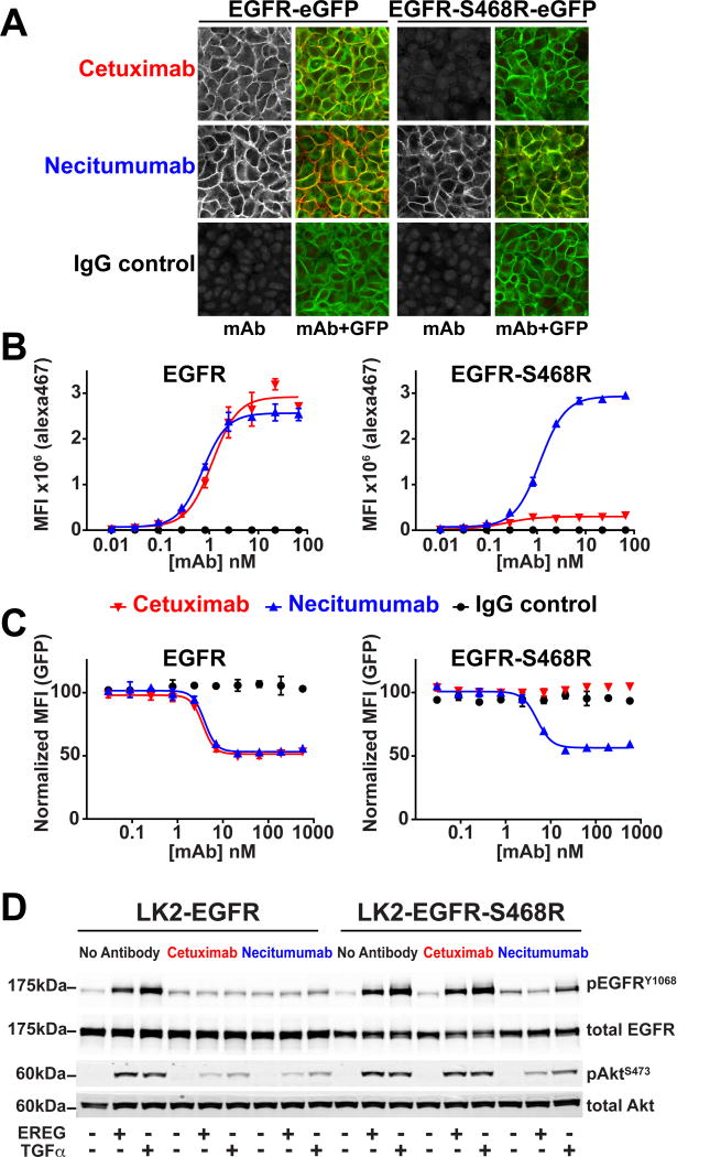
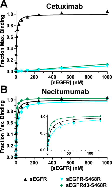
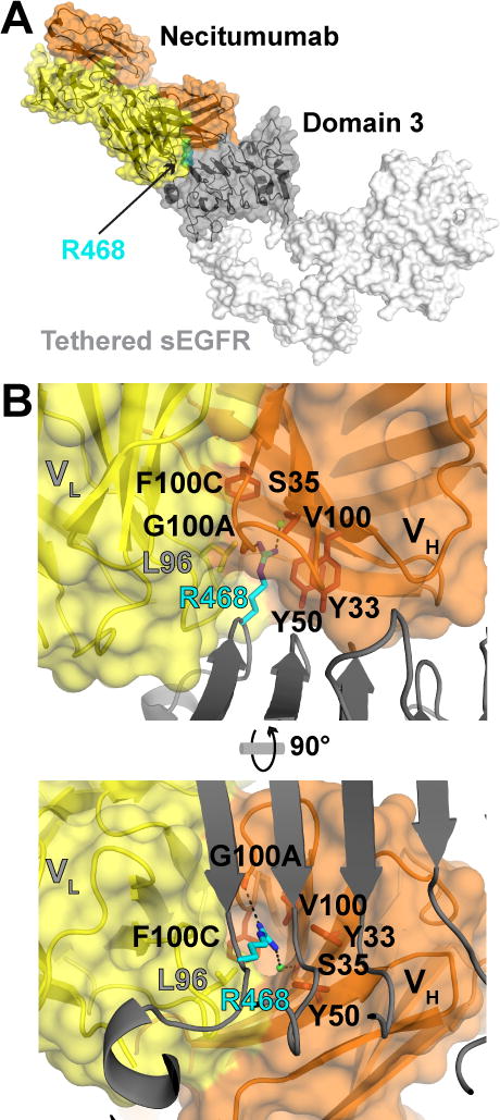
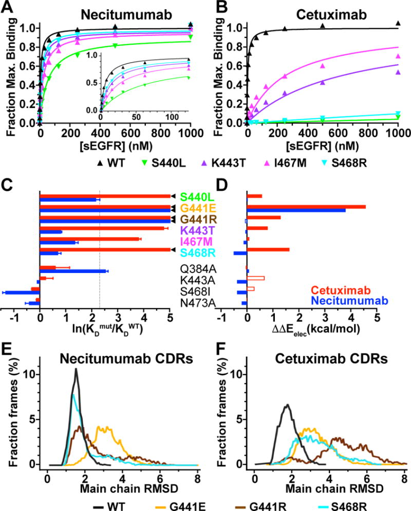
Acquired resistance to cetuximab, an antibody that targets the EGFR, impacts clinical benefit in head and neck, and colorectal cancers. One of the mechanisms of resistance to cetuximab is the acquisition of mutations that map to the cetuximab epitope on EGFR and prevent drug binding. We find that necitumumab, another FDA-approved EGFR antibody, can bind to EGFR that harbors the most common cetuximab-resistant substitution, S468R (or S492R, depending on the amino acid numbering system). We determined an X-ray crystal structure to 2.8 Å resolution of the necitumumab Fab bound to an S468R variant of EGFR domain III. The arginine is accommodated in a large, preexisting cavity in the necitumumab paratope. We predict that this paratope shape will be permissive to other epitope substitutions, and show that necitumumab binds to most cetuximab- and panitumumab-resistant EGFR variants. We find that a simple computational approach can predict with high success which EGFR epitope substitutions abrogate antibody binding. This computational method will be valuable to determine whether necitumumab will bind to EGFR as new epitope resistance variants are identified. This method could also be useful for rapid evaluation of the effect on binding of alterations in other antibody/antigen interfaces. Together, these data suggest that necitumumab may be active in patients who are resistant to cetuximab or panitumumab through EGFR epitope mutation. Furthermore, our analysis leads us to speculate that antibodies with large paratope cavities may be less susceptible to resistance due to mutations mapping to the antigen epitope. Mol Cancer Ther; 17(2); 521-31. ©2017 AACR.


Figures

Figure 1
Figure 1. Necitumumab binds to and inhibits EGFR harboring the S468R cetuximab resistance mutation
A, confocal imaging of HELA cells expressing EGFR-eGFP and EGFR-S468R-eGFP stained with alexa647 labeled antibodies (left, gray scale). Right panels show an overlay of the alexa647 and GFP fluorescence. B, binding of the same antibodies to EGFR and EGFR-S468R expressing HELA cells analyzed by flow cytometry. The median fluorescence intensity (MFI) values are plotted over a concentration range from 0.01 to 67 nM of cetuximab (red), necitumumab (blue) or control IgG (black). There is negligible binding of cetuximab or necitumumab to parental HELA cells over this concentration range (Supplementary Fig. S1C). Plots are derived from three technical replicates, and are representative of data from three independent experiments. C, normalized GFP MFI signal for HELA cells expressing EGFR-eGFP or EGFR-S469R-eGFP following 48-hours pre-incubated with the indicated concentrations of cetuximab (red), necitumumab (blue), or IgG (black). MFI values are normalized to the EGFR-GFP signal with no antibody treatment. Plots are from two technical replicates and representative of data from two independent experiments. D, effect of antibody pre-treatment (1,000 nM) on EGFR activation in LK2-EGFR or LK2-EGFR-S468R cells. Cells were stimulated with EREG or TGFα (100 nM and 10 nM, respectively) and immunoblotted for total and phosphorylated EGFR and AKT (pEGFRY1068 and pAktS473).
Figure 2
Figure 2. Necitumumab binds with high affinity to sEGFR harboring the cetuximab resistance mutations S468R
A and B, SPR analysis of sEGFR, sEGFR-S468R and sEGFRd3-S468R binding to immobilized cetuximab (A) and necitumumab (B) Fabs. Normalized equilibrium SPR response plotted as a function of protein concentration were fit to a simple one-site Langmuir binding equation. Data are representative of at least three independent measurements. Mean KD values with standard deviations are reported in Supplementary Table S2.
Figure 3
Figure 3. Structure of the sEGFRd3-S468R/Fab11F8 complex
A, a transparent surface representation plus cartoon of the sEGFRd3-S468R/Fab11F8 structure with antibody orange (VH) and yellow (VL) and domain III dark gray. The R468 side chain is in sphere representation colored cyan. Domains I, II and IV that are not present in the structure are in white (from PDB 1YY9). B, orthogonal views of the necitumumab paratope, with VH and VL colored as in A, and domain III in gray cartoon. R468 (cyan) sits in a deep hydrophobic cavity between the VH and VL domains. Amino acids lining the cavity are shown in stick representation. Presumed direct and water mediated hydrogen bonds with R468 are indicated with dashed lines.
Figure 4
Figure 4. Comparison of necitumumab and cetuximab paratopes
A, surface representations of necitumumab and cetuximab (PDB 1YY9) paratopes in the same orientation as in Fig. 3B (right). The electrostatic potential, calculated using the adaptive Poisson-Boltzmann solver (APBS) (49), from −5 kT (red) to +5 kT (blue) is projected on to the surface. R468 (left) and S468 (right) are in cyan stick representation. B, the paratope surfaces are colored to highlight the position of the CDRs. VL CDRs are yellow, CDR H1 pale orange, CDR H2, orange and CDR H3 brown. Side chains from the CDR H3 of cetuximab that interact with side chains in the VL CDRs are show.
Figure 5
Figure 5. Experimental and computational evaluation of the effect of epitope substitutions on cetuximab and necitumumab binding to EGFR
A and B, SPR binding of indicated sEGFR variants to necitumumab (A) and cetuximab (B) Fabs, analyzed as described in the legend to Fig. 2. Mean KD values are in Supplementary Table S2. C, experimental change in binding due to the indicated substitution in sEGFR for cetuximab (red) and necitumumab (blue), plotted as the natural log of the ratio of the KD value for mutated EGFR to the KD value of WT; ln(KDmut/KDWT). KDmut/KDWT values from Table 1. ◄ indicates ln(KDmut/KDWT) >> 5. Dotted line is at KDmut/KDWT = 10. D, computed change in electrostatic binding energy for each mutated EGFR relative to WT; ΔΔEelec = ΔEelecmut - ΔEelecWT colored as in C. Open bars indicated incorrect predictions (see text and Table 1). E, dynamics of necitumumab CDRs during three independent 200 ns MD simulations when bound to WT (black), G441E (yellow), G441R (brown) and S468R (cyan) EGFR. Structures from 6000 frames were aligned to frame 1 using main chain of domain III. Binned (0.07 Å) RMSD values for main chain CDR atoms, expressed as a percentage of the total number of frames, are plotted as a function of RMSD. Mean (median) of the RMSD cumulative distributions are; WT: 1.54 (1.5) Å, S468R: 1.96 (1.64) Å, G441R: 2.64 (2.17) Å, and G441E: 3.19 (3.14) Å. Differences in mean for WT and G441 substitutions are significant (P<0.001). F, the same analysis as in E for the complexes with cetuximab. Mean (median) values; WT: 1.86 (1.81) Å, S468R: 3.33 (3.19) Å, G441R: 4.71 (4.73) Å, and G441E: 3.31 (3.21) Å. Differences for WT is significant (P<0.001).

References

    1. Arteaga CL, Engelman JA. ERBB receptors: from oncogene discovery to basic science to mechanism-based cancer therapeutics. Cancer Cell. 2014;25:282–303. - PMC - PubMed
    1. Roskoski R., Jr The ErbB/HER family of protein-tyrosine kinases and cancer. Pharmacol Res. 2014;79:34–74. - PubMed
    1. Veronese ML, O’Dwyer PJ. Monoclonal antibodies in the treatment of colorectal cancer. Eur J Cancer. 2004;40:1292–301. - PubMed
    1. Lee MS, Kopetz S. Current and Future Approaches to Target the Epidermal Growth Factor Receptor and Its Downstream Signaling in Metastatic Colorectal Cancer. Clin Colorectal Cancer. 2015;14:203–18. - PubMed
    1. Okines A, Cunningham D, Chau I. Targeting the human EGFR family in esophagogastric cancer. Nat Rev Clin Oncol. 2011;8:492–503. - PubMed

Publication types

MeSH terms