. 2017 Nov 20;8(1):1623.
doi: 10.1038/s41467-017-01743-6.

Annexin A4 and A6 induce membrane curvature and constriction during cell membrane repair

Affiliations

Annexin A4 and A6 induce membrane curvature and constriction during cell membrane repair

Theresa Louise Boye et al. Nat Commun. .

Abstract

Efficient cell membrane repair mechanisms are essential for maintaining membrane integrity and thus for cell life. Here we show that the Ca2+- and phospholipid-binding proteins annexin A4 and A6 are involved in plasma membrane repair and needed for rapid closure of micron-size holes. We demonstrate that annexin A4 binds to artificial membranes and generates curvature force initiated from free edges, whereas annexin A6 induces constriction force. In cells, plasma membrane injury and Ca2+ influx recruit annexin A4 to the vicinity of membrane wound edges where its homo-trimerization leads to membrane curvature near the edges. We propose that curvature force is utilized together with annexin A6-mediated constriction force to pull the wound edges together for eventual fusion. We show that annexin A4 can counteract various plasma membrane disruptions including holes of several micrometers indicating that induction of curvature force around wound edges is an early key event in cell membrane repair.


Conflict of interest statement

The authors declare no competing financial interests.

Figures

Fig. 1
Fig. 1
ANXA4 counteracts plasma membrane injuries triggered by detergent, hypo-osmotic stress, and heat shock. a Immunofluorescence images showing endogenous ANXA4 protein localization in HeLa cells (white arrows) exposed to digitonin (20 µg/ml, 15 min), hypo-osmotic stress (25 mOsm/l, 10 min) or 47 °C heat shock (5 min) (scale bar, 20 µm). Plasma membrane integrity assays in cells overexpressing ANXA4-RFP or Ctrl-RFP and b treated with digitonin (20 µg/ml) in HeLa cells, or c MCF7 cells exposed to hypo-osmotic stress (25 mOsm/l) and assayed with impermeable Hoechst-33258. Also, see Supplementary Fig. 1. d Images showing FM1-43 dye uptake in MCF7 cells exposed to hypo-osmotic stress and e corresponding intensity plot. f Plasma membrane integrity upon 55 °C heat shock in MCF7 cells expressing ANXA4 wild type, or g Ca2+-binding-deficient mutants, h mutant lacking N terminus (aa 4-13) (ΔN). i Residues mutated in the Ca2Mut/Ca4Mut and the TrimMut ANXA4 mutants are indicated with yellow and pink sticks, respectively, on the crystal structure of ANXA4 (PDB entry code: 2zoc) displayed in transparent light blue cartoon with bound calcium ions as blue spheres. j ANXA4 (light blue) is structurally aligned on a subunit of homotrimer observed in the crystal structure of ANXA5 (transparent gray, PDB entry code: 1a8a) and the residues mutated in the ANXA4- TrimMut mutant displayed as pink spheres. The two structures are aligned to a root-mean-square deviation (RMSD) value of 0.942 Å over 302 Cα atoms (PyMOL Molecular Graphics System, Version 1.7.6.4, Schrödinger, LLC). k Membrane integrity upon 55 °C heat shock in MCF7 cells expressing ANXA4-TrimMut as compared to wtANXA4 and control. Means of three independent experiments measuring >40 cells for each condition. Error bars represent S.D. for three independent experiments. P values based on t-test: *P ≤ 0.05, **P ≤ 0.01, ***P ≤ 0.001
Fig. 2
Fig. 2
ANXA4 is needed for repair in MCF7 cells. a Immunoblot of lysates from MCF7A4-CRISPR cells with disrupted ANXA4 gene expression. Hsc70 served as internal control for equal loading. b Plasma membrane repair kinetics upon laser injury measured by membrane impermeable FM1-43 dye influx in b CRISPR clones and in c MCF7A4-CRISPR (#1) cells expressing wild-type ANXA4-RFP, ANXA4 TrimMut-RFP or Ctrl-RFP. Error bars represent S.D. for at least eight independent cells per condition. P values based on t-test: *P ≤ 0.05, **P ≤ 0.01. d Sequential 3D images of MCF7A4---CRISPR (#1) with reintroduced ANXA4-RFP and exposed to a large injury by shooting through the cell by laser (injury site marked by white arrow). ANXA4-RFP translocation to wound edges and constriction is marked by yellow arrow (also, see Supplementary Movie 1). e Example of ANXA4-RFP-induced curvature and funnel formation (yellow arrow) created from the bottom of the plasma membrane after laser injury. f Trimer-deficient ANXA4-RFP mutant (A4-TrimMut) failed to induce curvature upon laser injury (see also Supplementary Movie 1)
Fig. 3
Fig. 3
ANXA4 induces membrane curvature in supported membranes. a Schematic of supported membrane model composed of primary and secondary membranes. Non-vesicular membrane patches with open edges were used in the subsequent experiments (POPC/POPS, 9:1 molar ratio). b Sequential images before and after addition of ANXA4-GFP protein to a membrane patch stained with DiD in the presence of Ca2+ (last image GFP channel) (also, see Supplementary Movie 2). c Similar experiment as in b but without POPS in the supported membrane. d, e Mutants of ANXA4 proteins including d Ca4Mut or e TrimMut. Note: TrimMut exhibits >10 times slower rolling activity. f Fluorescence and matching AFM image of membrane patch after completed rolling induced by ANXA4 and g AFM-topography graph of two colliding membrane rolls. h Schematic of ANXA4-induced membrane rolling including a numeric estimate of the number of turns N and layer spacing d. i Calculation of time constant (τ) for membrane rolling and corresponding data for ANXA4. ANXA4-GFP shows similar rolling activity (data not shown). j Rolling time constants for ANXA4 and A4 ΔN, respectively. Error bars represent S.D. for three–five independent experiments. P values based on t-test: *P ≤ 0.05, ***P ≤ 0.001
Fig. 4
Fig. 4
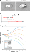
Biophysical model for ANXA4-induced curvature during repair. a Initial and curved state of a circular membrane hole. The solid area inside the solid circle is the region contributing to the change in curvature elastic energy between the initial and curved states. b Profile of the neck and definitions: initial hole radius r 0, the neck angle Δ and curvature radius B. c Energy difference ΔH as a function of Δ for varying values of the initial hole size. When r 0 is below a critical hole radius r 0* there is a minimum in ΔH corresponding to a energetically favored neck angle Δmin. In the plot r 0 ≈ 800 nm. For r 0 > r 0*, ΔH decreases monotonically with Δ meaning that continuous rolling is favored (also, see Supplementary Notes 1 and 2 for detailed description)
Fig. 5
Fig. 5
ANXA6 triggers in-plane constriction of membrane edges and shrinkage of supported membrane islands. a Sequential images before and after addition of ANXA6 protein to a membrane patch stained with DiD in the absence of Ca2+ or b in the presence of Ca2+. Note that ANXA6 induces in-plane folding of membrane edges (lower panel, magnified image) as well as larger out-of-plane folds (white arrows). c Fluorescence and matching AFM images of membrane patch after completed ANXA6 folding. Lower panel: corresponding AFM-topography graph. d Model for ANXA6-triggered local membrane constriction by cross bridging of adjacent membrane via the two core domains
Fig. 6
Fig. 6
ANXA6 is recruited to wound edges and required for repair. a Representative sequential 3D images of MCF cell showing the localization of ANXA6-GFP and ANXA4-RFP in response to laser injury (white arrows indicate injury site). Cells were injured by shooting through the cell to obtain a hole with clear edge. A putative repair cap defining where the edges have fused is visible in the middle (yellow arrow). b Immunoblot showing ANXA6 protein levels in CRISP Ctrl and MCF7A4-CRISPR (#1) cells or in similar cells treated with ANXA6 or Ctrl siRNA for 48 h. c Cell membrane repair kinetics in ANXA6 siRNA-treated cells compared to Ctrl siRNA-treated cells upon laser injury measured by membrane impermeable FM1-43 dye influx in MCF7Ctrl CRISPR or d MCF7A4--CRISPR (#1). Error bars represent S.D. for at least seven independent cells per condition. P values based on t-test: *P ≤ 0.05, **P ≤ 0.01. **P ≤ 0.001
Fig. 7
Fig. 7
Proposed model for plasma membrane repair initiated by ANXA4 and ANXA6. a Binding of ANXA6 to wound edges induces local in-plane folding, which contracts the hole edges. b In uninjured cells, ANXA6 and ANXA4 are distributed uniformly as monomers in the cytoplasm. Upon local plasma membrane injury, Ca2+ influx results in recruitment of ANXA6 and ANXA4 to the membrane wound edges. ANXA6 initiates constriction of hole edges and may also cross-bridge patch vesicles translocated to the injury site. ANXA4 self-assembles into trimers that induce local out-of-plane curvature. The combined forces of constriction and curvature accelerate wound closure eventually leading to fusion of membrane edges. Vesicles recruited and fused to the wound edges contribute to repair by reducing wound size. The curvature structure triggered by ANXA4 can take several shapes including I round or II funnel-shaped or a more flat structure (not shown). Subsequent repair steps include local actin polymerization to fully restore the membrane after injury

Similar articles

Cited by

References

    1. Cooper ST, McNeil PL. Membrane repair: mechanisms and pathophysiology. Physiol. Rev. 2015;95:1205–1240. doi: 10.1152/physrev.00037.2014. - DOI - PMC - PubMed
    1. Idone V, Tam C, Andrews NW. Two-way traffic on the road to plasma membrane repair. Trends Cell Biol. 2008;18:552–559. doi: 10.1016/j.tcb.2008.09.001. - DOI - PMC - PubMed
    1. Bement WM, Mandato CA, Kirsch MN. Wound-induced assembly and closure of an actomyosin purse string in Xenopus oocytes. Curr. Biol. 1999;9:579–587. doi: 10.1016/S0960-9822(99)80261-9. - DOI - PubMed
    1. Idone V, et al. Repair of injured plasma membrane by rapid Ca2+-dependent endocytosis. J. Cell Biol. 2008;180:905–914. doi: 10.1083/jcb.200708010. - DOI - PMC - PubMed
    1. Jimenez AJ, et al. ESCRT machinery is required for plasma membrane repair. Science. 2014;343:1247136. doi: 10.1126/science.1247136. - DOI - PubMed

Publication types