. 2017 Nov 20;7(1):15826.
doi: 10.1038/s41598-017-15870-z.

Treatment of Myocardial Infarction with Gene-modified Mesenchymal Stem Cells in a Small Molecular Hydrogel

Affiliations

Treatment of Myocardial Infarction with Gene-modified Mesenchymal Stem Cells in a Small Molecular Hydrogel

Zhiye Wu et al. Sci Rep. .

Abstract

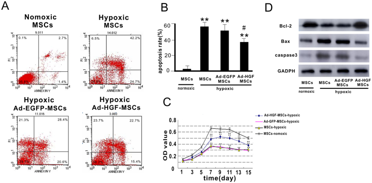
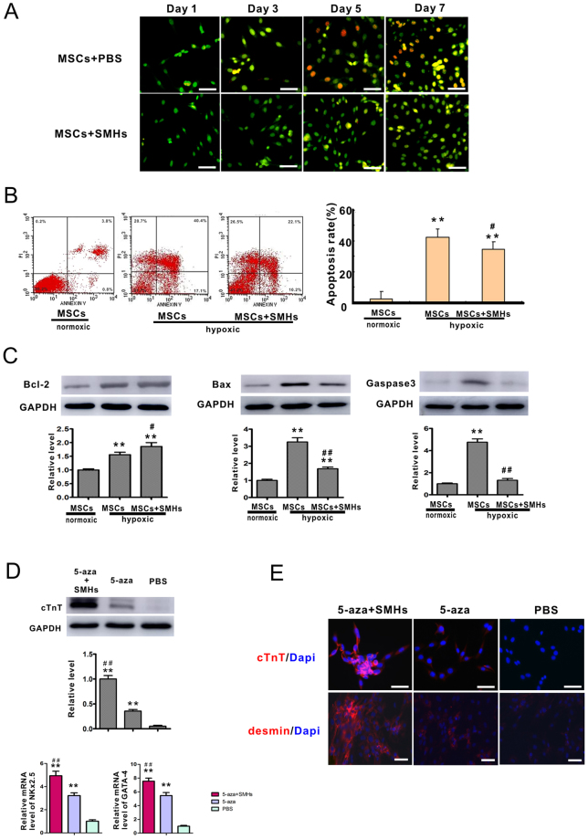
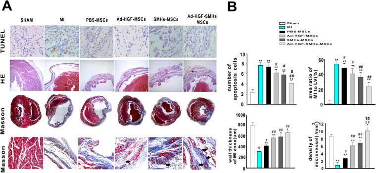
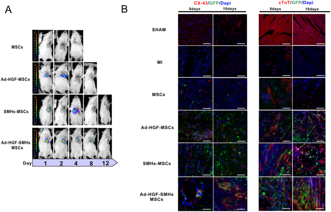
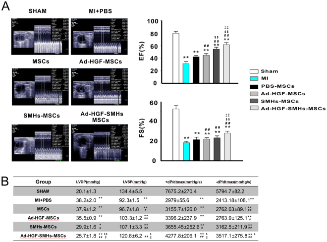
The effect of transplanted rat mesenchymal stem cells (MSCs) can be reduced by extracellular microenvironment in myocardial infarction (MI). We tested a novel small-molecular hydrogel (SMH) on whether it could provide a scaffold for hepatocyte growth factor (HGF)-modified MSCs and alleviate ventricular remodeling while preserving cardiac function after MI. Overexpression of HGF in MSCs increased Bcl-2 and reduced Bax and caspase-3 levels in response to hypoxia in vitro. Immunocytochemistry demonstrated that cardiac troponin (cTnT), desmin and connexin 43 expression were significantly enhanced in the 5-azacytidine (5-aza) with SMH group compared with the 5-aza only group in vitro and in vivo. Bioluminescent imaging indicated that retention and survival of transplanted cells was highest when MSCs transfected with adenovirus (ad-HGF) were injected with SMH. Heart function and structure improvement were confirmed by echocardiography and histology in the Ad-HGF-SMHs-MSCs group compared to other groups. Our study showed that: HGF alleviated cell apoptosis and promoted MSC growth. SMHs improved stem cell adhesion, survival and myocardial cell differentiation after MSC transplantation. SMHs combined with modified MSCs significantly decreased the scar area and improved cardiac function.


Conflict of interest statement

The authors declare that they have no competing interests.

Figures

Figure 1
Figure 1
Infection of HGF into MSCs and the effect on MSC growth and apoptosis after. 12 hours of hypoxia. (A) Flow cytometry analysis of Annexin V/PI was carried out in MSCs. (B) Apoptosis rate was calculated according to Annexin V/PI flow cytometry analysis. (C) MTT curve of MSCs infected with Ad-HGF or vector in normoxic or hypoxic conditions. (D) Representative Western blot and level of Bcl-2, Bax and caspase-3. Data are mean ± SEM. **P < 0.01 vs. MSCs-normoxic, # P < 0.5, ## P < 0.1 vs. Ad-EGFP-MSCs-hypoxic. n = 3, One-way ANOVA, then followed by post-hoc Tukey’s test for multiple comparisons.
Figure 2
Figure 2
The effect of SMHs on MSC proliferation, survival and differentiation. (A) MSCs cultured in SMHs or normal medium were visualized by AO/EB staining, magnification:400x. (B) Annexin V/ PI flow cytometry analysis with MSCs cultured with SMHs in hypoxic or normoxic conditions. Data are mean ± SEM. *P < 0.05 vs. MSCs-normoxic, # P < 0.05 vs. MSCs-hypoxic,n = 3. (C) Western blot and level of Bcl-2, Bax and caspase-3 in each group. Levels of proteins were quantified by densitometry and normalized against GAPDH, data are mean ± SEM. *P < 0.05 vs. MSCs-normoxic, # P < 0.05 vs. MSCs-hypoxic, all the experiments were repeated three times then selected the representative result for each protein level. (D) Representative western blot and the level of cTnT; Level of NKx2.5 and GATA-4 were detected by qPCR. Data are mean ± SEM. **P < 0.05 vs. PBS; ## P < 0.01 vs. 5-AZA. n = 3, Levels of proteins and mRNA were quantified by densitometry and normalized against GAPDH, One-way ANOVA, then followed by post-hoc Tukey’s test for multiple comparisons. (E) SMHs affected MSC differentiation as shown by red fluorescence in photomicrographs: cTnT, and desmin, Blue: DAPI-stained nuclei, magnification:400x.
Figure 3
Figure 3
Immunohistochemistry staining in vivo. (A) In the peri-infarcted area apoptotic cells were detected and calculated by TUNEL assay. Photomicrographs of the LV tissue sections stained by H&E and Masson staining exhibited increased micro-vessel density, ratio of the MI area to LV%, and wall thickness in the MI zone. Magnification: 400x. (B) The graph shows the statistics of apoptotic cells, density of micro-vessel and ratio of the MI area to LV% as well as wall thickness in the MI zone. Data are mean ± SEM. *or **P < 0.05 or 0.01 vs. Sham; # or ## P < 0.05 or 0.01 vs. group MI. $or $$ P < 0.05 or < 0.01 vs. SMH-MSCs and Ad-HGF-MSCs, n = 9~10.
Figure 4
Figure 4
Immunofluorescence and bioluminescent imaging in vivo. (A) Retention and survival signals in transplanted cells were monitored by non-invasive bioluminescent imaging technology. (B) MSC retention and survival induced by 5-aza in each treatment group was measured by green fluorescence; The expression of cTnT and Cx43 in the 8days and 16days section were displayed as red fluorescence, Blue fluorescence: DAPI-stained nuclei n = 6. Magnification: 400x.
Figure 5
Figure 5
Improvement of cardiac function. (A) Representative pictures of echocardiography in each group and EF(%) and FS(%) statistics from bar graphs. (B) Table for cardiac hemodynamic monitoring. Data are mean ± SEM. *P < 0.05, **P < 0.01 vs. Sham; # P < 0.05, ## P < 0.01 vs. MI + PBS; $ P < 0.05, $$ P < 0.01 vs. Ad-HGF-MSCs; P < 0.05 vs. SMHs-MSCs; n = 9~10. One-way ANOVA, then followed by post-hoc Tukey’s test for multiple comparisons.

References

    1. Ishikawa K, et al. Stem cell factor gene transfer improves cardiac function after myocardial infarction in swine. Circulation. Heart failure. 2015;8:167–174. doi: 10.1161/CIRCHEARTFAILURE.114.001711. - DOI - PMC - PubMed
    1. Meyer GP, et al. Intracoronary bone marrow cell transfer after myocardial infarction: eighteen months’ follow-up data from the randomized, controlled BOOST (BOne marrOw transfer to enhance ST-elevation infarct regeneration) trial. Circulation. 2006;113:1287–1294. doi: 10.1161/CIRCULATIONAHA.105.575118. - DOI - PubMed
    1. Korf-Klingebiel M, et al. Bone marrow cells are a rich source of growth factors and cytokines: implications for cell therapy trials after myocardial infarction. European heart journal. 2008;29:2851–2858. doi: 10.1093/eurheartj/ehn456. - DOI - PubMed
    1. Behfar A, Terzic A. Stem cell in the rough: repair quotient mined out of a bone marrow niche. Circulation research. 2014;115:814–816. doi: 10.1161/CIRCRESAHA.114.305075. - DOI - PubMed
    1. Trachtenberg B, et al. Rationale and design of the Transendocardial Injection of Autologous Human Cells (bone marrow or mesenchymal) in Chronic Ischemic Left Ventricular Dysfunction and Heart Failure Secondary to Myocardial Infarction (TAC-HFT) trial: A randomized, double-blind, placebo-controlled study of safety and efficacy. American heart journal. 2011;161:487–493. doi: 10.1016/j.ahj.2010.11.024. - DOI - PubMed

MeSH terms