. 2017 Oct 19:17:222-231.
doi: 10.1016/j.nicl.2017.10.019. eCollection 2018.

Disruption of default mode network dynamics in acute and chronic pain states

Affiliations

Disruption of default mode network dynamics in acute and chronic pain states

Z Alshelh et al. Neuroimage Clin. .

Abstract

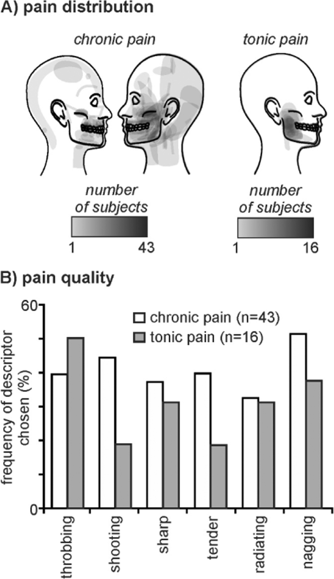
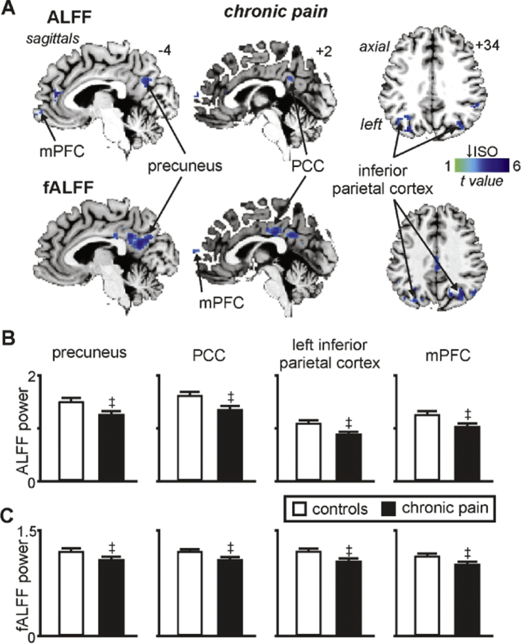
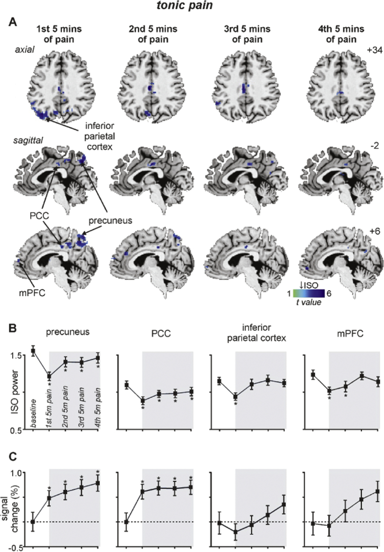
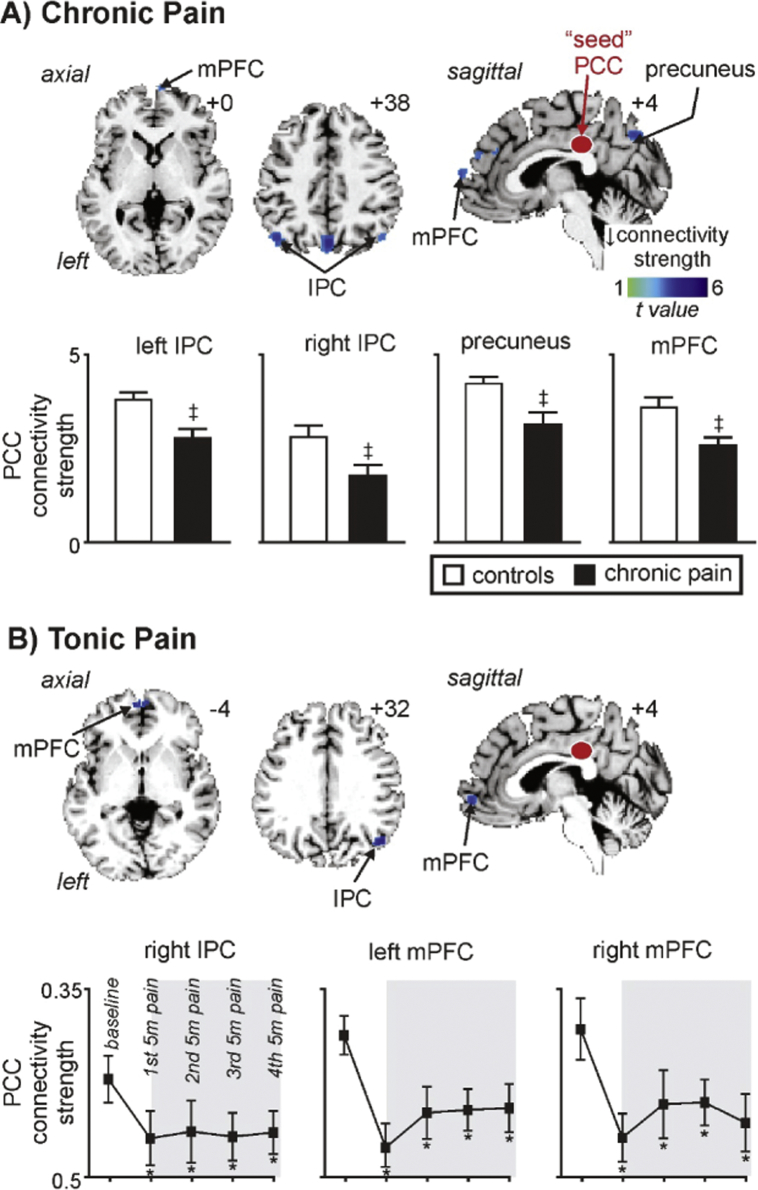
It has been proposed that pain competes with other attention-demanding stimuli for cognitive resources, and many chronic pain patients display significant attention and mental flexibility deficits. These alterations may result from disruptions in the functioning of the default mode network (DMN) which plays a critical role in attention, memory, prospection and self-processing, and recent investigations have found alterations in DMN function in multiple chronic pain conditions. Whilst it has been proposed that these DMN alterations are a characteristic of pain that is chronic in nature, we recently reported altered oscillatory activity in the DMN during an acute, 5 minute noxious stimulus in healthy control subjects. We therefore hypothesize that altered DMN activity patterns will not be restricted to those in chronic pain but instead will also occur in healthy individuals during tonic noxious stimuli. We used functional magnetic resonance imaging to measure resting state infra-slow oscillatory activity and functional connectivity in patients with chronic orofacial pain at rest and in healthy controls during a 20-minute tonic pain stimulus. We found decreases in oscillatory activity in key regions of the DMN in patients with chronic pain, as well as in healthy controls during tonic pain in addition to changes in functional connectivity between the posterior cingulate cortex and areas of the DMN in both groups. The results show that similar alterations in DMN function occur in healthy individuals during acute noxious stimuli as well as in individuals with chronic pain. These DMN changes may reflect the presence of pain per se and may underlie alterations in attentional processes that occur in the presence of pain.

Keywords: ALFF, amplitude of low-frequency fluctuations; Attention; Chronic orofacial pain; DMN, default mode network; Functional magnetic resonance imaging; IPC, inferior parietal cortex; ISO, infra-slow oscillations; PCC, posterior cingulate cortex; Posterior cingulate cortex; Precuneus; Prefrontal cortex; fALFF, fractional amplitude of low-frequency fluctuations; mPFC, medial prefrontal cortex.


Figures

Fig. 1
Fig. 1
Pain distribution and quality of pain in chronic pain subjects and in individuals during tonic pain. A) Individual pain distribution patterns in 43 chronic pain subjects and 16 healthy controls during experimentally induced tonic pain. B) Frequency (percentage of subjects) of descriptors chosen from the McGill Pain Questionnaire to describe the on-going pain in chronic pain subjects (white bars) and in healthy controls during tonic pain (grey bars).
Fig. 2
Fig. 2
The default mode network in controls, chronic pain and tonic pain groups as defined by an independent component analysis. Areas displaying significant signal intensity covariation are indicated by the hot colour scale and overlaid onto an individual's T1-weighted anatomical image set. The default mode network consists of the precuneus, posterior cingulate cortex (PCC), inferior parietal cortex and the medial prefrontal cortex (mPFC). Slice locations in Montreal Neurological Institute space are indicated at the top right of each slice in the top panel.
Fig. 3
Fig. 3
Significant changes in infra-slow oscillatory power (ISO: 0.03–0.06 Hz) in individuals with chronic pain. A) Regions where chronic pain subjects have significantly reduced amplitude of low-frequency fluctuations (ALFF) and fractional ALFF (fALFF) compared with controls within the default mode network. Power reductions are indicated by the cool colour scale and overlaid onto an individual's T1-weighted anatomical image set. Note the decreases within the precuneus, posterior cingulate cortex (PCC), inferior parietal cortex and medial prefrontal cortex (mPFC). Slice locations in Montreal Neurological Institute space are indicated at the top right of each slice in the top panel. B) Plots of mean ± SEM ALFF and C) mean ± SEM fALFF in controls and chronic pain groups for significant clusters. ‡ significant differences derived from the voxel-by-voxel random effects analysis (p < 0.05, false discovery rate corrected).
Fig. 4
Fig. 4
Significant changes in infra-slow oscillatory power (ISO: 0.03–0.06 Hz) during tonic pain. A) Regions of the default mode network where the amplitude of low-frequency fluctuations (ALFF) are significantly reduced during the 1st, 2nd, 3rd and 4th 5  minute periods of tonic pain compared with a baseline period. Power reductions are indicated by the cool colour scale and overlaid onto an individual's T1-weighted anatomical image set. Note the decreases within the precuneus, posterior cingulate cortex (PCC), inferior parietal cortex and medial prefrontal cortex (mPFC). Slice locations in Montreal Neurological Institute space are indicated at the top right of each slice in panel A. B) Plots of mean ± SEM ALFF during baseline and the four 5  minute pain periods for significant clusters. C) Plots of mean ± SEM % signal intensity changes relative to baseline for the four 5  minute pain periods for clusters with significantly reduced ALFF power. Note that only the precuneus and PCC display both reduced ALFF and increased signal intensity changes during all of the tonic pain periods. The grey shading indicates the tonic pain period. * p < 0.05, 2-tailed paired t-test.
Fig. 5
Fig. 5
Significant changes in posterior cingulate cortex (PCC) functional connectivity in chronic and tonic pain groups. A) Regions where PCC connectivity strength within the default mode network is significantly reduced in chronic pain subjects compared with controls. Connectivity strength reductions are indicated by the cool colour scale and overlaid onto an individual's T1-weighted anatomical image set. Note decreases in the precuneus, inferior parietal cortex (IPC) and medial prefrontal cortex (mPFC). Slice locations in Montreal Neurological Institute space are indicated at the top right of each slice. Plots of mean ± SEM PCC connectivity strength are also shown. ‡ significant differences derived from the voxel-by-voxel random effects analysis (p < 0.05, false discovery rate corrected). B) Regions where PCC connectivity strength is significantly reduced during tonic pain compared with the baseline period. Connectivity strength reductions are indicated by the cool colour scale and overlaid onto an individual's T1-weighted anatomical image set. Note the decreases within the IPC and mPFC. Plots of mean ± SEM PCC connectivity strength are also shown for the baseline and each of the 5 min tonic pain periods. The grey shading indicates the tonic pain period. * p < 0.05, 2-tailed paired t-test.

References

    1. Alshelh Z., Di Pietro F., Youssef A.M., Reeves J.M., Macey P.M., Vickers E.R., Peck C.C., Murray G.M., Henderson L.A. Chronic neuropathic pain: It's about the rhythm. J. Neurosci. 2016;36:1008–1018. - PMC - PubMed
    1. Baliki M.N., Geha P.Y., Apkarian A.V., Chialvo D.R. Beyond feeling: chronic pain hurts the brain, disrupting the default-mode network dynamics. J. Neurosci. 2008;28:1398–1403. - PMC - PubMed
    1. Baliki M.N., Mansour A.R., Baria A.T., Apkarian A.V. Functional reorganization of the default mode network across chronic pain conditions. PLoS One. 2014;9 - PMC - PubMed
    1. Bantick S.J., Wise R.G., Ploghaus A., Clare S., Smith S.M., Tracey I. Imaging how attention modulates pain in humans using functional MRI. Brain. 2002;125:310–319. - PubMed
    1. Brown G.D., Yamada S., Sejnowski T.J. Independent component analysis at the neural cocktail party. Trends Neurosci. 2001;24:54–63. - PubMed