

Using simultaneous PET/MRI to compare the accuracy of diagnosing frontotemporal dementia by arterial spin labelling MRI and FDG-PET
- PMID: 29159053
- PMCID: PMC5683801
- DOI: 10.1016/j.nicl.2017.10.033
Using simultaneous PET/MRI to compare the accuracy of diagnosing frontotemporal dementia by arterial spin labelling MRI and FDG-PET
Abstract
Purpose: The clinical utility of FDG-PET in diagnosing frontotemporal dementia (FTD) has been well demonstrated over the past decades. On the contrary, the diagnostic value of arterial spin labelling (ASL) MRI - a relatively new technique - in clinical diagnosis of FTD has yet to be confirmed. Using simultaneous PET/MRI, we evaluated the diagnostic performance of ASL in identifying pathological abnormalities in FTD (FTD) to determine whether ASL can provide similar diagnostic value as FDG-PET.
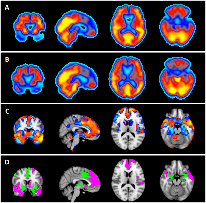
Methods: ASL and FDG-PET images were compared in 10 patients with FTD and 10 healthy older adults. Qualitative and quantitative measures of diagnostic equivalency were used to determine the diagnostic utility of ASL compared to FDG-PET. Sensitivity, specificity, and inter-rater reliability were calculated for each modality from scores of subjective visual ratings and from analysis of regional mean values in thirteen a priori regions of interest (ROI). To determine the extent of concordance between modalities in each patient, individual statistical maps generated from comparison of each patient to controls were compared between modalities using the Jaccard similarity index (JI).
Results: Visual assessments revealed lower sensitivity, specificity and inter-rater reliability for ASL (66.67%/62.12%/0.2) compared to FDG-PET (88.43%/90.91%/0.61). Across all regions, ASL performed lower than FDG-PET in discriminating patients from controls (areas under the receiver operating curve: ASL = 0.75 and FDG-PET = 0.87). In all patients, ASL identified patterns of reduced perfusion consistent with FTD, but areas of hypometabolism exceeded hypoperfused areas (group-mean JI = 0.30 ± 0.22).
Conclusion: This pilot study demonstrated that ASL can detect similar spatial patterns of abnormalities in individual FTD patients compared to FDG-PET, but its sensitivity and specificity for discriminant diagnosis of a patient from healthy individuals remained unmatched to FDG-PET. Further studies at the individual level are required to confirm the clinical role of ASL in FTD management.
Keywords: Arterial spin labelling MRI; FDG-PET; Frontotemporal dementia; Hybrid PET/MRI.
Figures


References
-
- Albert, M., DeCarli, C.S., DeKosky, S.T., de Leon, M.J., Foster, N.L., Fox, N.C., Frank, R., Frackowiak, R.S., Jack, C.R., Jagust, W.J., Knopman, D.S., Morris, J.C., Petersen, R.C., Reiman, E., Scheltens, P., Small, G., Soininen, H., Thal, L., Wahlund, L.-O., Thies, W., Weiner, M., Khachaturian, Z., 2005. The use of MRI and PET for clinical diagnosis of dementia and investigation of cognitive impairment: a consensus report. Alzheimer's Assoc. Neuroimaging Work Gr. Consens. Rep. 1–15.
-
- Alsop, D.C., Detre, J.A., Golay, X., Günther, M., Hendrikse, J., Hernandez-Garcia, L., Lu, H., MacIntosh, B.J., Parkes, L.M., Smits, M., van Osch, M.J.P., Wang, D.J.J., Wong, E.C., Zaharchuk, G., 2015. Recommended implementation of arterial spin-labeled perfusion MRI for clinical applications: a consensus of the ISMRM perfusion study group and the European consortium for ASL in dementia. Magn. Reson. Med. 73, 102–116. doi:https://doi.org/10.1002/mrm.25197. - DOI - PMC - PubMed
-
- Anazodo U.C., Thiessen J.D., Ssali T., Mandel J., Günther M., Butler J., Pavlosky W., Prato F.S., Thompson R.T., St. Lawrence K.S. Feasibility of simultaneous whole-brain imaging on an integrated PET-MRI system using an enhanced 2-point Dixon attenuation correction method. Front. Neurosci. 2015;8:1–11. - PMC - PubMed
-
- Ashburner J., Friston K.J. Unified segmentation. NeuroImage. 2005;26:839–851. - PubMed
-
- Aslan, S., Xu, F., Wang, P.L., Uh, J., Yezhuvath, U.S., van Osch, M., Lu, H., 2010. Estimation of labeling efficiency in pseudocontinuous arterial spin labeling. Magn. Reson. Med. 63, 765–71. doi:https://doi.org/10.1002/mrm.22245. - DOI - PMC - PubMed
MeSH terms
Substances
LinkOut - more resources
Full Text Sources
Other Literature Sources
Medical
Miscellaneous
