

Linked color imaging (LCI), a novel image-enhanced endoscopy technology, emphasizes the color of early gastric cancer
- PMID: 29159276
- PMCID: PMC5634856
- DOI: 10.1055/s-0043-117881
Linked color imaging (LCI), a novel image-enhanced endoscopy technology, emphasizes the color of early gastric cancer
Abstract
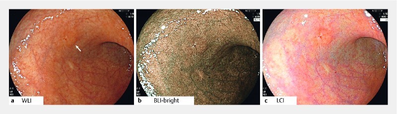
Background and study aims: Linked color imaging (LCI) and blue laser imaging (BLI) are novel image-enhanced endoscopy technologies with strong, unique color enhancement. We investigated the efficacy of LCI and BLI-bright compared to conventional white light imaging (WLI) by measuring the color difference between early gastric cancer lesions and the surrounding mucosa.
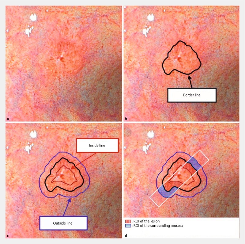
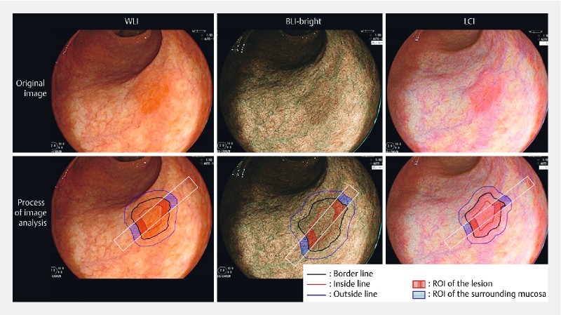
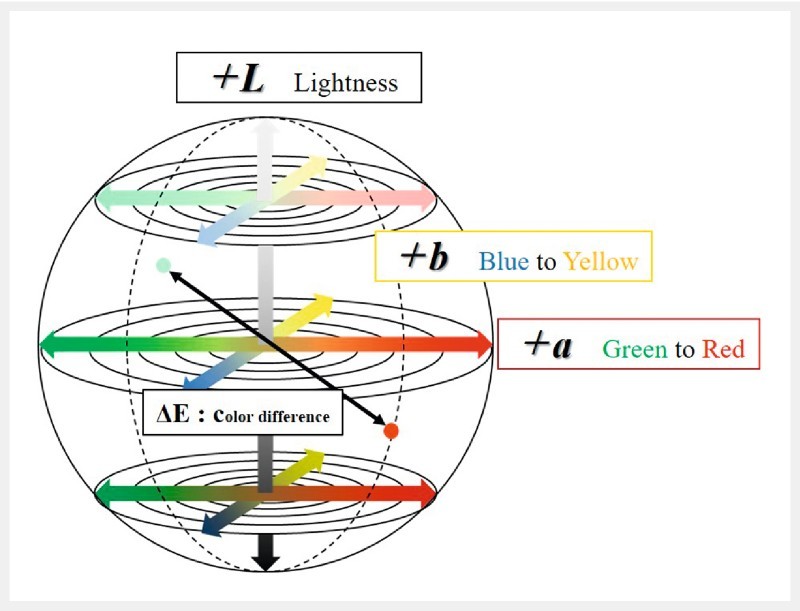
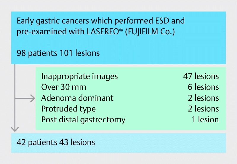
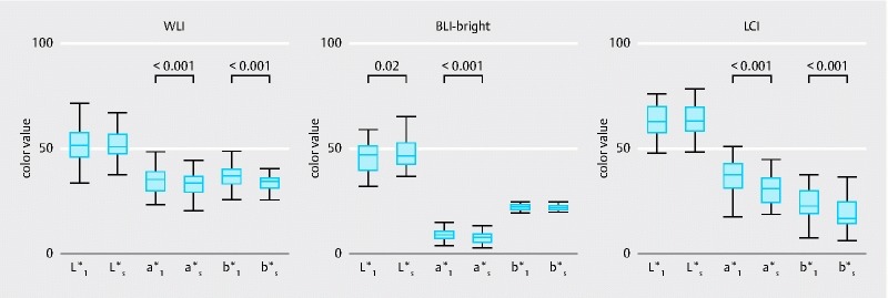
Patients and methods: Images of early gastric cancer scheduled for endoscopic submucosal dissection were captured by LCI, BLI-bright, and WLI under the same conditions. Color values of the lesion and surrounding mucosa were defined as the average of the color value in each region of interest. Color differences between the lesion and surrounding mucosa (ΔE) were examined in each mode. The color value was assessed using the CIE L*a*b* color space (CIE: Commission Internationale d'Eclairage).
Results: We collected images of 43 lesions from 42 patients. Average ΔE values with LCI, BLI-bright, and WLI were 11.02, 5.04, and 5.99, respectively. The ΔE was significantly higher with LCI than with WLI ( P < 0.001). Limited to cases of small ΔE with WLI, the ΔE was approximately 3 times higher with LCI than with WLI (7.18 vs. 2.25). The ΔE with LCI was larger when the surrounding mucosa had severe intestinal metaplasia ( P = 0.04). The average color value of a lesion and the surrounding mucosa differed. This value did not have a sufficient cut-off point between the lesion and surrounding mucosa to distinguish them, even with LCI.
Conclusion: LCI had a larger ΔE than WLI. It may allow easy recognition and early detection of gastric cancer, even for inexperienced endoscopists.
Conflict of interest statement
Figures
Similar articles
-
Linked-color Imaging May Help Improve the Visibility of Superficial Barrett's Esophageal Adenocarcinoma by Increasing the Color Difference.Intern Med. 2021;60(21):3351-3358. doi: 10.2169/internalmedicine.6674-20. Epub 2021 Nov 1. Intern Med. 2021. PMID: 34719622 Free PMC article.
-
Linked Color Imaging for Stomach.Diagnostics (Basel). 2023 Jan 27;13(3):467. doi: 10.3390/diagnostics13030467. Diagnostics (Basel). 2023. PMID: 36766572 Free PMC article. Review.
-
Utility of linked color imaging for endoscopic diagnosis of early gastric cancer.World J Gastroenterol. 2019 Mar 14;25(10):1248-1258. doi: 10.3748/wjg.v25.i10.1248. World J Gastroenterol. 2019. PMID: 30886507 Free PMC article.
-
[Clinical application of LASEREO endoscopic system in early gastric cancer].Zhonghua Nei Ke Za Zhi. 2022 Mar 1;61(3):310-316. doi: 10.3760/cma.j.cn112138-20210328-00246. Zhonghua Nei Ke Za Zhi. 2022. PMID: 35263973 Chinese.
-
Current status and future perspective of linked color imaging for gastric cancer screening: a literature review.J Gastroenterol. 2023 Jan;58(1):1-13. doi: 10.1007/s00535-022-01934-z. Epub 2022 Oct 26. J Gastroenterol. 2023. PMID: 36287268 Free PMC article. Review.
Cited by
-
Bile pigment in small-bowel water content may reflect bowel habits: a retrospective analysis of a capsule endoscopy imaging series.BMC Gastroenterol. 2020 Jul 23;20(1):237. doi: 10.1186/s12876-020-01382-0. BMC Gastroenterol. 2020. PMID: 32703159 Free PMC article.
-
Linked color imaging improves the endoscopic visibility of gastric mucosal cancers.Endosc Int Open. 2019 Feb;7(2):E164-E170. doi: 10.1055/a-0733-7086. Epub 2019 Jan 18. Endosc Int Open. 2019. PMID: 30705948 Free PMC article.
-
Linked-color Imaging May Help Improve the Visibility of Superficial Barrett's Esophageal Adenocarcinoma by Increasing the Color Difference.Intern Med. 2021;60(21):3351-3358. doi: 10.2169/internalmedicine.6674-20. Epub 2021 Nov 1. Intern Med. 2021. PMID: 34719622 Free PMC article.
-
Linked Color Imaging for Stomach.Diagnostics (Basel). 2023 Jan 27;13(3):467. doi: 10.3390/diagnostics13030467. Diagnostics (Basel). 2023. PMID: 36766572 Free PMC article. Review.
-
Additional Thirty Seconds Observation with Linked Color Imaging Improves Detection of Missed Polyps in the Right-Sided Colon.Gastroenterol Res Pract. 2018 Jul 8;2018:5059834. doi: 10.1155/2018/5059834. eCollection 2018. Gastroenterol Res Pract. 2018. PMID: 30116263 Free PMC article.
References
-
- Stewart B W, Wild C P. Lyon: World Health Organization; 2014. World cancer report 2014.
-
- Nieminen A, Kokkola A, Yla-Liedenpohja J et al.Early gastric cancer: clinical characteristics and results of surgery. Dig Surg. 2009;26:378–383. - PubMed
-
- Nam S Y, Choi I J, Park K W et al.Effect of repeated endoscopic screening on the incidence and treatment of gastric cancer in health screenees. Eur J Gastroenterol Hepatol. 2009;21:855–860. - PubMed
LinkOut - more resources
Full Text Sources
Other Literature Sources