. 2017 Oct 5:7:67-75.
doi: 10.1016/j.omto.2017.10.001. eCollection 2017 Dec 15.

Insertion of the Type-I IFN Decoy Receptor B18R in a miRNA-Tagged Semliki Forest Virus Improves Oncolytic Capacity but Results in Neurotoxicity

Affiliations

Insertion of the Type-I IFN Decoy Receptor B18R in a miRNA-Tagged Semliki Forest Virus Improves Oncolytic Capacity but Results in Neurotoxicity

Tina Sarén et al. Mol Ther Oncolytics. .

Abstract

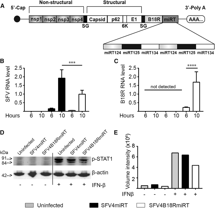
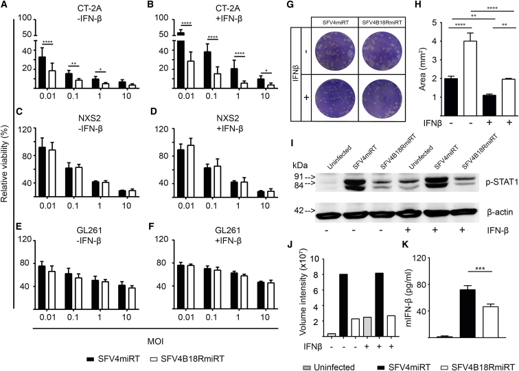
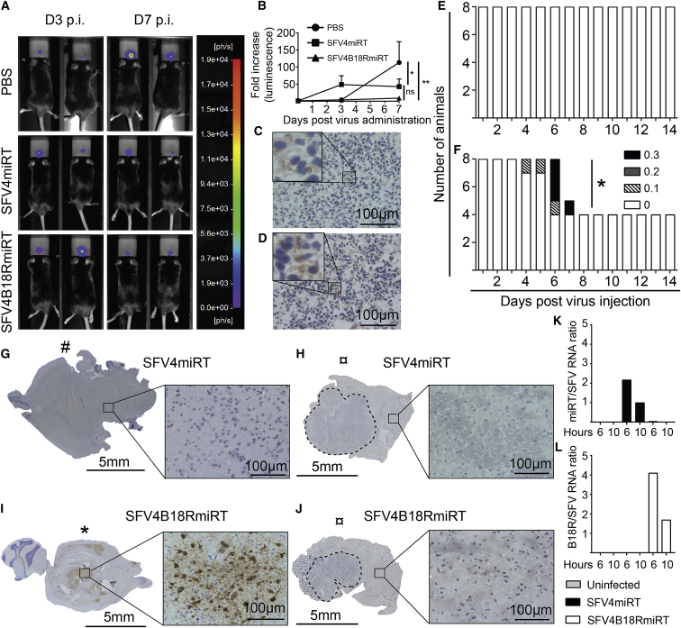
Oncolytic Semliki Forest virus (SFV) has been suggested as a potential candidate for the treatment of glioblastoma and neuroblastoma. However, the oncolytic capacity of SFV is restricted by the anti-viral type-I interferon (IFN) response. The aim of this study was to increase the oncolytic capacity of a microRNA target tagged SFV against glioblastoma by arming it with the Vaccinia-virus-encoded type-I IFN decoy receptor B18R (SFV4B18RmiRT) to neutralize type-I IFN response. Expression of B18R by SFV4B18RmiRT aided neutralization of IFN-β, which was shown by reduced STAT-1 phosphorylation and improved virus spread in plaque assays. B18R expression by SFV4 increased its oncolytic capacity in vitro against murine glioblastoma (CT-2A), regardless of the presence of exogenous IFN-β. Both SFV4B18RmiRT and SFV4miRT treatments controlled tumor growth in mice with syngeneic orthotopic gliomablastoma (CT-2A). However, treatment with SFV4B18RmiRT induced severe neurological symptoms in some mice because of virus replication in the healthy brain. Neither neurotoxicity nor virus replication in the brain was observed when SFV4miRT was administered. In summary, our results indicate that the oncolytic capacity of SFV4 was improved in vitro and in vivo by incorporation of B18R, but neurotoxicity of the virus was increased, possibly due to loss of microRNA targets.

Keywords: B18R; SFV; Semliki Forest virus; glioblastoma; type I interferon.


Figures

Figure 1
Figure 1
Functional Analysis of B18R Expressed by SFV4B18RmiRT (A) Schematic representation of the SFV construct. B18R was inserted downstream of the structural genes under the control of a subgenomic promoter (SG). Target sequences for miR-124, miR-125 and miR-134 (in duplicates) were inserted in the 3′ UTR. (B and C) CT-2A cells were infected (MOI 10) with SFV4miRT or SFV4B18RmiRT, and RNA was extracted from infected cells after 6 and 10 hr. Uninfected cells were used as controls. Real-time PCR was used to determine SFV nsP3 (B) and B18R (C) RNA levels. RNA levels were normalized toward GAPDH. Values are shown as mean ± SEM and were compared by one-way ANOVA with Sidak post hoc test (n = 3, ****p < 0.0001, ***p < 0.001). (D) Western blot for detection of p-STAT1 (Tyr701) in CT-2A cells. Supernatants were collected from BHK21 cells infected (MOI 10) with SFV4miRT or SFV4B18RmiRT overnight. Exogenous mIFN-β (200 pg/mL) was added to the supernatant for 60 min to allow for B18R in the SFV4B18RmiRT supernatant to neutralize IFN-β. Supernatants were then added to CT-2A cells for 30 min, after which p-STAT1 was detected using western blot to determine IFN-β activity and thereby indirect B18R neutralizing capacity. (E) Quantification of p-STAT1 (both isoforms) from 1 day presented as volume intensity normalized to β-actin. The blot was analyzed in ImageLab software.
Figure 2
Figure 2
Presence of B18R Increases Oncolytic SFV Capacity and Viral Spread In Vitro (A–F) The ability of SFV4miRT and SFV4B18RmiRT to kill CT-2A (A and B), NXS2 (C and D), and GL261 (E and F) cells was assessed in the absence (A, C, and E) or presence (B, D, and F) of exogenous mIFN-β (10 pg/mL). Cells were infected with an MOI gradient ranging from 0.01 to 10, and cell viability was determined using the alamarBlue assay 48 or 72 hr after infection. The experiment was repeated at least twice with internal triplicates. Values are shown as mean ± SEM and were compared by two-way ANOVA with Holm-Sidak post hoc test (****p < 0.0001, **p < 0.01, *p < 0.05). (G and H) Viral spread was assessed in a plaque assay by infecting NXS2 cells at MOI 0.005 with or without exogenous mIFN-β (10 pg/mL). Plaques were detected using crystal violet 3 days p.i. (G), and the size (mm2) was determined using ImageJ. Values are shown as mean ± SEM (H) and were compared by one-way ANOVA with Tukey post hoc test (****p < 0.0001, **p < 0.01). (I and J) The amount of p-STAT1 (Tyr701) in CT-2A cells upon SFV4miRT or SFV4B18RmiRT infection was determined using western blot (I) and quantified (J). CT-2A cells were infected at MOI 10 for 6 hr and left untreated or treated with mIFN-β (50 pg/mL) for 30 min. One representative blot is shown from duplicate experiments. (J) Quantification of p-STAT1 (both isoforms) presented as volume intensity normalized to β-actin. The blots were analyzed in ImageLab. (K) mIFN-β quantification in the supernatant of CT-2A cells infected with SFV4miRT or SFV4B18RmiRT at MOI 10 after overnight incubation using ELISA. The experiment was repeated at least two times with internal duplicates. Means were compared by one-way ANOVA with Tukey post hoc test (***p < 0.001).
Figure 3
Figure 3
Therapeutic Efficacy and Neurotoxicity of SFV4B18RmiRT To determine the therapeutic efficacy of SFV4B18RmiRT in vivo, CT-2A/fLuc cells were injected orthotopically in the brain of C57BL/6NRj mice. 11 days later, when tumors were visible by in vivo bioluminescence imaging, mice were injected (i.p.) either with PBS (n = 5), SFV4miRT (n = 8), or SFV4B18RmiRT (n = 8) at 1 × 107 PFU. (A) Representative pictures of mice with CT-2A/fLuc tumors 3 and 7 days after virus administration. (B) Quantification of tumor growth (fold increase in luminescence) after virus administration (PBS n = 5, SFV4miRT n = 8, and SFV4B18RmiRT n = 5). Values are shown as mean ± SEM and were compared by two-way ANOVA with Tukey post hoc test (**p < 0.01; *p < 0.05; not significant [n.s.], p > 0.05). (C and D) Immunohistochemical staining for SFV proteins in the tumor area in a mice brain injected with SFV4miRT (C) or SFV4B18RmiRT (D) (optical magnification 40X, inset 4x digital magnification). (E and F) Scoring of neurological symptoms in mice injected with SFV4miRT (n = 8) (E) or SFV4B18RmiRT (n = 8) (F). The symptoms were scored in a scale ranging from 0 (no symptoms) to 0.3 (severe or accumulation of several mild symptoms). When reaching a score of 0.3, mice were sacrificed. (G–J) Brains were excised from virus-injected mice and stained for SFV proteins in the non-tumor area of the brain in mice injected with SFV4miRT that were sacrificed early (#, control) (G) or late (H) due to tumor burden (¤) and mice injected with SFV4B18RmiRT sacrificed early (I) due to virus-related symptoms (*) or late (J) due to tumor burden (¤). (K and L) SFV (nsp), B18R, and miRT RNA levels in SFV4miRT- and SFVB18RmiRT-infected CT-2A cells were determined by qPCR. Cells were infected at MOI 10, and RNA was extracted 6 and 10 hr p.i. RNA levels were normalized toward GAPDH. miRT (K) and B18R (L) expression level was related to viral replication (SFV RNA level). #, sacrificed as control; *, sacrificed due to virus-related symptoms (neurotoxicity); ¤, sacrificed due to tumor burden.

References

    1. Alifieris C., Trafalis D.T. Glioblastoma multiforme: pathogenesis and treatment. Pharmacol. Ther. 2015;152:63–82. - PubMed
    1. Furnari F.B., Fenton T., Bachoo R.M., Mukasa A., Stommel J.M., Stegh A., Hahn W.C., Ligon K.L., Louis D.N., Brennan C. Malignant astrocytic glioma: genetics, biology, and paths to treatment. Genes Dev. 2007;21:2683–2710. - PubMed
    1. Ostrom Q.T., Gittleman H., Fulop J., Liu M., Blanda R., Kromer C., Wolinsky Y., Kruchko C., Barnholtz-Sloan J.S. CBTRUS statistical report: primary brain and central nervous system tumors diagnosed in the United States in 2008-2012. Neuro. Oncol. 2015;17(Suppl 4):iv1–iv62. - PMC - PubMed
    1. Kaufman H.L., Kohlhapp F.J., Zloza A. Oncolytic viruses: a new class of immunotherapy drugs. Nat. Rev. Drug Discov. 2015;14:642–662. - PMC - PubMed
    1. Fazakerley J.K. Pathogenesis of Semliki Forest virus encephalitis. J. Neurovirol. 2002;8(Suppl 2):66–74. - PubMed