. 2018 Aug;596(15):3149-3169.
doi: 10.1113/JP275001. Epub 2018 Jan 4.

Acute intermittent hypoxia with concurrent hypercapnia evokes P2X and TRPV1 receptor-dependent sensory long-term facilitation in naïve carotid bodies

Affiliations

Acute intermittent hypoxia with concurrent hypercapnia evokes P2X and TRPV1 receptor-dependent sensory long-term facilitation in naïve carotid bodies

Arijit Roy et al. J Physiol. 2018 Aug.

Abstract

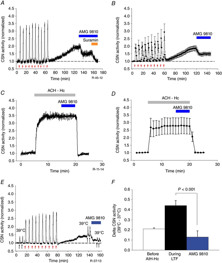
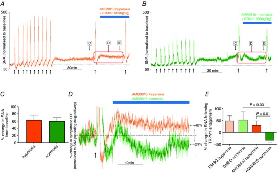
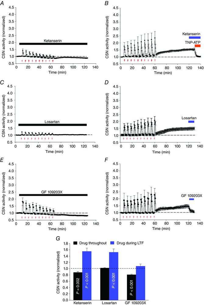
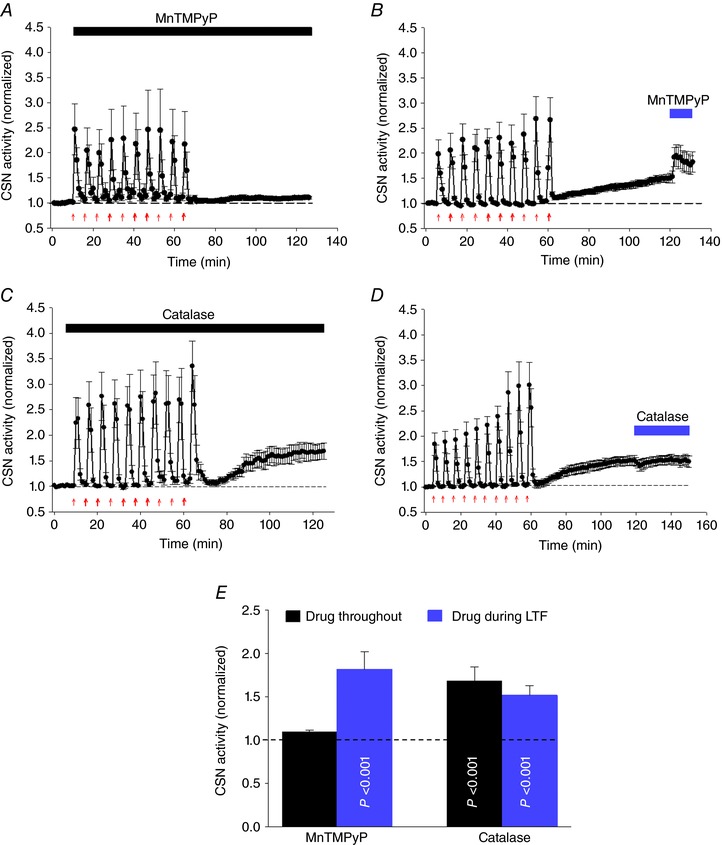
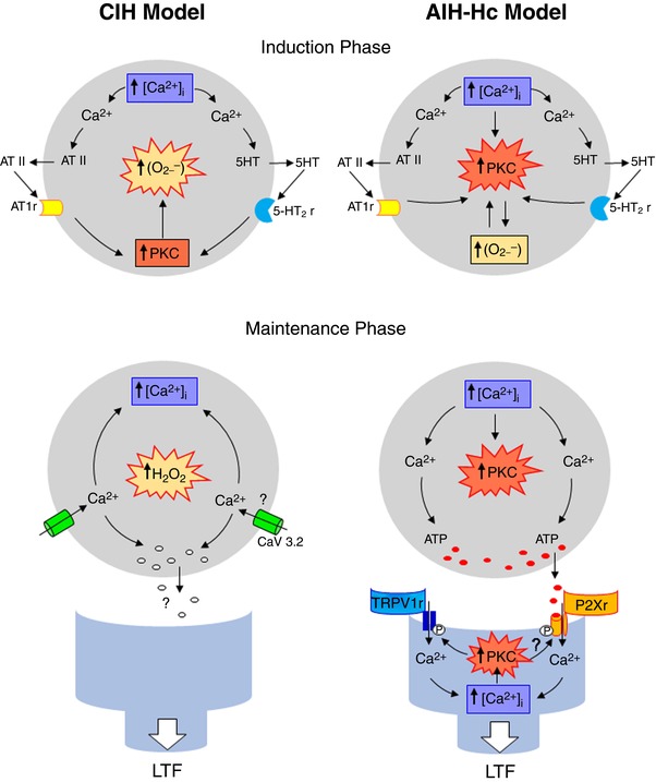
Key points: Activity-dependent plasticity can be induced in carotid body (CB) chemosensory afferents without chronic intermittent hypoxia (CIH) preconditioning by acute intermittent hypoxia coincident with bouts of hypercapnia (AIH-Hc). Several properties of this acute plasticity are shared with CIH-dependent sensory long-term facilitation (LTF) in that induction is dependent on 5-HT, angiotensin II, protein kinase C and reactive oxygen species. Several properties differ from CIH-dependent sensory LTF; H2 O2 appears to play no part in induction, whereas maintenance requires purinergic P2X2/3 receptor activation and is dependent on transient receptor potential vanilloid type 1 (TRPV1) receptor sensitization. Because P2X2/3 and TRPV1 receptors are located in carotid sinus nerve (CSN) terminals but not presynaptic glomus cells, a primary site of the acute AIH-Hc induced sensory LTF appears to be postsynaptic. Our results obtained in vivo suggest a role for TRPV1-dependent CB activity in acute sympathetic LTF. We propose that P2X-TRPV1-receptor-dependent sensory LTF may constitute an important early mechanism linking sleep apnoea with hypertension and/or cardiovascular disease.

Abstract: Apnoeas constitute an acute existential threat to neonates and adults. In large part, this threat is detected by the carotid bodies, which are the primary peripheral chemoreceptors, and is combatted by arousal and acute cardiorespiratory responses, including increased sympathetic output. Similar responses occur with repeated apnoeas but they continue beyond the last apnoea and can persist for hours [i.e. ventilatory and sympathetic long-term facilitation (LTF)]. These long-term effects may be adaptive during acute episodic apnoea, although they may prolong hypertension causing chronic cardiovascular impairment. We report a novel mechanism of acute carotid body (CB) plasticity (sensory LTF) induced by repeated apnoea-like stimuli [i.e. acute intermittent hypoxia coincident with bouts of hypercapnia (AIH-Hc)]. This plasticity did not require chronic intermittent hypoxia preconditioning, was dependent on P2X receptors and protein kinase C, and involved heat-sensitive transient receptor potential vanilloid type 1 (TRPV1) receptors. Reactive oxygen species (O2 ·¯) were involved in initiating plasticity only; no evidence was found for H2 O2 involvement. Angiotensin II and 5-HT receptor antagonists, losartan and ketanserin, severely reduced CB responses to individual hypoxic-hypercapnic challenges and prevented the induction of sensory LTF but, if applied after AIH-Hc, failed to reduce plasticity-associated activity. Conversely, TRPV1 receptor antagonism had no effect on responses to individual hypoxic-hypercapnic challenges but reduced plasticity-associated activity by ∼50%. Further, TRPV1 receptor antagonism in vivo reduced sympathetic LTF caused by AIH-Hc, although only if the CBs were functional. These data demonstrate a new mechanism of CB plasticity and suggest P2X-TRPV1-dependent sensory LTF as a novel target for pharmacological intervention in some forms of neurogenic hypertension associated with recurrent apnoeas.

Keywords: carotid body; hypoxia-hypercapnia; sensory long term facilitation.


Figures

Figure 1
Figure 1. AIH‐Hc causes sensory LTF in ex vivo CB from naïve rat
Examples of rectified CB sensory activity (AB) from three preparations exposed to ten 1 min bouts of hypoxia‐hypercapnia (red arrows; each interspersed with 5 min of normoxia‐normocapnia). Insets represent action potentials from a single unit. During AIH‐Hc (i.e. the induction phase), most preparations had augmenting responses with each bout (A, recirculating effluent; B, non‐recirculating effluent). We observed a small number of preparations with decrementing responses, C, after AIH‐Hc (i.e. during the maintenance phase), CSN activity increased in (A) and (B) (indicative of sensory LTF) but not in (C) (no sensory LTF). D, magnitude of CSN activity 60 min after the last hypoxia‐hypercapnia bout (n = 63). In total, 57 preparations demonstrated sensory LTF (blue); in the remaining six, sensory LTF was absent (grey). E, scatter plot showing the relationship between the change in CSN activity between the first and last (10th) bout of hypoxia‐hypercapnia compared to the CSN activity at 60 min after the last bout. Only preparations in which responses increased with bouts demonstrated sensory LTF (blue). However, the degree of augmentation did not predict sensory LTF magnitude (P = 0.06; r 2 = 0.02). Note that CSN activity is normalized to the baseline preceding the first bout (dashed line). [Color figure can be viewed at http://wileyonlinelibrary.com]
Figure 2
Figure 2. Sensory LTF in ex vivo CBs from naïve rats depends on stimulus pattern and strength
Average integrated CSN activity in response to: (A) no gaseous challenge (control; open circles) or AIH‐Hc (red arrows; closed circles); (B) 10 bouts of hypoxia (PO2 = 60 Torr; blue arrows) with concurrent sustained Hc (SHc) (PCO2 = 50 Torr; blue horizontal bar); (C) 10 bouts of hypoxia (PO2 = 40 Torr; green arrows); (D) 10 bouts of Hc (PCO2 = 60 Torr; orange arrows); and (E) 10 min of acute continuous hypoxia‐hypercapnia (ACH‐Hc) (PO2 = 40 Torr & PCO2 = 60 Torr; grey bar). F, summary of CSN activity 60 min after the last stimuli. Note that concurrent hypercapnia, which enhances the acute response of the CB to hypoxia, was necessary to induce sensory LTF. * P < 0.05 compared to baseline (dashed line) (one‐way ANOVA). All data are the mean ± SEM (n = 6 per group). rc, recirculated effluent; wrc, without recirculated effluent.
Figure 3
Figure 3. AIH‐Hc induced sensory LTF sensitizes responses to hypoxia and heat of CBs from naïve rats
A and C, examples of integrated CSN responses to acute hypoxia (PO2 = 60 Torr for 3 min; black horizontal bars) and temperature challenge (37°C to 39°C for 1 min; black horizontal bars), respectively, before and 60 min after 10 episodes of AIH‐Hc (red arrows). Note the increased hypoxic and temperature responses during the maintenance phase of sensory LTF compared to control (pre‐AIH‐Hc). B and D, summary of the effects of hypoxia and temperature challenges, respectively, before and during sensory LTF. Data are the mean ± SEM (n = 5 per group). Comparison between delta changes before and during sensory LTF. N, normoxia; H, hypoxia.
Figure 4
Figure 4. AIH‐Hc induced sensory LTF involves P2X receptors and increases sensitivity to ATP
A and B, average integrated CSN activity showing sensory LTF induced by AIH‐Hc is eliminated by broad spectrum purinergic P2X receptor antagonist suramin and P2X 2/3 receptor‐specific antagonist TNP‐ATP, respectively (orange bars; mean ± SEM; n = 6). C, integrated CSN activity from one preparation showing the response to a bolus of ATP (1 ml of 100 μM ATP for 1 min; black arrows) in hyperoxia (PO2 ∼ 500 Torr; blue areas) before and during sensory LTF. Prior to AIH‐Hc, hyperoxia, by silencing the CB, possibly eliminates presynaptic ATP release from glomus cells (their primary neurotransmitter) and all but abolishes CSN activity; this is transiently reversed by exogenous ATP, presumably acting on postsynaptic P2X receptors. After AIH‐Hc, hyperoxia reduces but does not abolish CSN activity and the ATP response is enhanced. D, summary of CSN activity in hyperoxia and with ATP challenges, before and during sensory LTF (mean ± SEM; n = 5); Comparison between delta changes with ATP, before and during sensory LTF. Hpx, hyperoxia. E, CB sensory responses to repetitive applications of 100 μm ATP (ten 1 min boluses at 5 min intervals; as indicated by the red arrows) produced mild sLTF; application of AMG 9810 (10 μm) partially suppressed sLTF (Fig. 5). F, individual scatter plot (n = 6) and average data of sensory LTF 60 min after the last ATP pulse (mean ± SEM; n = 6; * P < 0.05 compared to baeline). Hpx, hyperoxia.
Figure 5
Figure 5. AIH‐Hc induced sensory LTF involves heat‐sensitive TRPV1 receptors and increases CB heat sensitivity
A, integrated CSN activity from one preparation showing ∼50% of sensory LTF induced by AIH‐Hc is inhibited by TRPV1 receptor antagonist AMG 9810 (10 μm; blue bar). The facilitated activity that remained was completely suppressed by suramin (100 μm; orange bar). B, average integrated CSN activity showing the effect of AMG9810 on sensory LTF (mean ± SEM; n = 5). C and D, integrated CSN activity from one preparation and average data (mean ± SEM; n = 6; P = 0.97) showing that AMG 9810 had no effect on the response to 10 min of acute continuous hypoxia‐hypercapnia (ACH‐Hc) (grey bar). E, integrated CSN activity from one preparation showing responses to temperature challenges (37 to 39°C for 1 min each; black arrows) before, 60 min after 10 episodes of AIH‐Hc (red arrows) and, subsequently, with partial inhibition of sensory LTF with TRPV1 antagonist AMG9810 (10 μm). F, summary of CSN responses following temperature challenge. Data are the mean ± SEM; n = 5.
Figure 6
Figure 6. Effect of TRPV1 antagonism on AIH‐Hc‐induced sympathetic LTF in naïve rats in vivo
A and B, integrated sympathetic responses to AIH‐Hc from two preparations with hyperoxic (orange trace) and normoxic (green trace) backgrounds, respectively. Note the sustained increase in sympathetic activity (indicative of sympathetic LTF) following the last bout of hypoxia‐hypercapnia. C, the magnitude of sympathetic LTF 60 min post‐AIH‐Hc was not different between preparations given normoxia or hyperoxia backgrounds. D, TRPV1 antagonist AMG9810 reduced sympathetic LTF, in preparations with a normoxic background (green) when the CB was functional but not in preparations with a hyperoxic background (orange). E, summary data showing percentage change in sympathetic activity following DMSO or AMG9810 administration in preparations with normoxic and hyperoxic backgrounds. AMG9810 significantly decreased sympathetic LTF under normoxic conditions but had no effect under hyperoxic conditions. Data are the mean ± SEM (n = 6).
Figure 7
Figure 7. Involvement of 5‐HT2 and AT1 receptors, and PKC activation in AIH‐Hc induced sensory LTF in CBs from naïve rats
Average integrated CSN activity (mean ± SEM; n = 5) showing that the anti‐hypertensive drugs ketanserin (A, 1 μm; 5‐HT2 receptor antagonist) and losartan (C, 3 μm; AT1 receptor antagonist) applied throughout the experiment diminished responses to hypoxia‐hypercapnia bouts (red arrows) and prevented sensory LTF. B and D, ketanserin and losartan, respectively, had no effect during the maintenance phase (i.e. once sensory LTF was established), whereas TNP‐ATP (P2X 2/3 receptor‐specific antagonist) completely abolished sensory LTF (as shown in Fig. 4 B). E, PKC inhibitor (GF 109203X; 10 μm) applied throughout the experiment also diminished responses to hypoxia‐hypercapnia bouts and prevented sensory LTF. F, when applied during the maintenance phase, GF 109203X potently inhibited sensory LTF (mean ± SEM; n = 5). G, summary data; P values were determined by comparing CSN activities with drugs applied throughout/after sensory LTF and the basal responses before AIH‐Hc (dashed line). Data are the mean ± SEM; n = 5).
Figure 8
Figure 8. ROS (O2·¯), but not H2O2, is involved in sensory LTF induction evoked by AIH‐Hc in naïve CB
A and B, average integrated CSN activity (mean ± SEM; n = 5) showing that ROS scavenger MnTMPyP (25 μm) applied throughout (red arrows) prevents sensory LTF but failed to suppress sensory LTF 60 min after the last hypoxia‐hypercapnia bout. C and D, average integrated CSN activity (mean ± SEM; n = 5) demonstrating the H2O2 scavenger PEG‐catalase (200 U ml−1) had no effect when applied either the induction or maintenance phase of sensory LTF. E, summary data of MnTMPyP and catalase applied throughout and after sensory LTF induction. P values were determined by comparing CSN activities with basal responses (dashed line) before AIH‐Hc stimuli. Data are the mean ± SEM; n = 5.
Figure 9
Figure 9. Schematic diagram comparing mechanisms of sensory LTF induction and maintenance by AIH requiring CIH pre‐conditioned and AIH‐Hc in CB from naïve rats
Although sensory LTF requiring CIH pre‐conditioning is largely dependent on O2. and subsequent production of H2O2, sensory LTF resulting from AIH with concurrent hypercapnia is largely dependent on PKC. Moreover, our novel data suggest that sensory LTF resulting from AIH with concurrent hypercapnia is P2X‐dependent, involves TRPV1 and is predominantly postsynaptic.

Comment in

References

    1. Bao G, Randhawa PM & Fletcher EC (1997). Acute blood pressure elevation during repetitive hypocapnic and eucapnic hypoxia in rats. J Appl Physiol 82, 1071–1078. - PubMed
    1. Bhave G, Hu H‐J, Glauner KS, Zhu W, Wang H, Brasier DJ, Oxford GS & Gereau RW (2003). Protein kinase C phosphorylation sensitizes but does not activate the capsaicin receptor transient receptor potential vanilloid 1 (TRPV1). Proc Natl Acad Sci USA 100, 12480–12485. - PMC - PubMed
    1. Caterina MJ, Schumacher MA, Tominaga M, Rosen TA, Levine JD & Julius D (1997). The capsaicin receptor: a heat‐activated ion channel in the pain pathway. Nature 389, 816–824. - PubMed
    1. Chin K, Hiras M, Kuriyama T, Fukui M, Kuno K, Sagawa Y & Ohi M (1997). Changes in the arterial PCO2 during a single night's sleep in patirnts with obstructive sleep apnea. Intern Med 36, 454–460. - PubMed
    1. Cummings KJ & Wilson RJA (2005). Time‐dependent modulation of carotid body afferent activity during and after intermittent hypoxia. Am J Physiol Regul Integr Comp Physiol 288, R1571–R1580. - PubMed

Publication types

LinkOut - more resources